Stomach, Abnormal Post Surgical Anatomy, Retained Fundus after Sleeve Gastrectomy
Stomach, Abnormal Post Surgical Anatomy, Retained Fundus after Sleeve Gastrectomy Video donated to the SAGES Video Atlas of Endoscopy by Eric M Pauli, MD Keyword(s): abnormal post surgical anatomy, retained fundus, SG, stomach
Retained Pill In Stomach
This video shows the endoscopic appeareance of a pill within the stomach. Video donated to the SAGES Video Atlas of Endoscopy by Eric M Pauli, MD Keyword(s): pill, stomach
Large Posterior Gastro Gastric Fistula after Gastric Bypass
This video shows the endoscopic view of a large, posterior-medial gastro-gastric fistula in a patient who had previously undergone a roux-en-Y gastric bypass for obesity. He had regained all of the weight he had previously lost. The fistula is seen posteriorly, and the gastro-jejunal anastomosis is seen in the distance, the scope traverses the anastomosis […]
Gastric Submucosal Hematoma
This video demonstrates a large submucosal gastric hematoma appreciated on retroflexed view in the fundus. Video donated to the SAGES Video Atlas of Endoscopy by Jose Martinez, J Andres Astudillo Keyword(s): fundus, Gastric Submucosal Hematoma, retroflexed view
Dialated Gastro-jejunal Anastomosis after Gastric Bypass – 2
This video shows a dilated gastro-jejunal anastomosis after a Roux-en-Y gastric bypass. The gastro-jejunal anastomosis was 40 mm by 45 mm as measured by the open snare. The patient has lost his sense of post-prandial satiety and had regained weight following the procedure. Donated to the SAGES Video Atlas of Endoscopy by Eric M Pauli, […]
Stomach, Abnormal Post Surgical Anatomy, Herniated Nissen Fundoplication
This video demonstrates a herniated and twisted wrap status post Nissen Fundoplication. Video donated to the SAGES Video Atlas of Endoscopy by Eric M Pauli, MD, Jeff Ponsky, MD Keyword(s): herniated Nissen fundoplication, post surgical anatomy, stomach, twisted wrap
Retained Fundus In A Paraesophageal Hernia after Sleeve Gastrectomy
This video shows a patient who had retained fundus during a sleeve gastrectomy due to the presence of the fundus within a paraesophageal hernia. The fundus was above the diaphragm at the time of the sleeve and was no resected. Video donated to the SAGES Video Atlas of Endoscopy by Eric M Pauli, MD Keyword(s): […]
Large Bleeding Gastro-jejunal Anastomotic Ulceration after Gastric Bypass
This video shows a large, deep gastro-jejunal anastomotic ulcer following a roux-en-y gastric bypass. There is a vissable vessel distally on the ulcer, and stigmata of bleeding (blood and small clot within the lumen) Video donated to the SAGES Video Atlas of Endoscopy by Eric M Pauli, MD Keyword(s): bleeding, blood clot, GJ anastomotic ulceration, […]
Gastric Polyps
This video demonstrates gastric polyps Video donated to the SAGES Video Atlas of Endoscopy by Jose Martinez, J Andres Astudillo Keyword(s): gastric polyps
Dialated Gastro-jejunal Anastomosis after Gastric Bypass
This video shows a dilated gastro-jejunal anastomosis after a Roux-en-Y gastric bypass. The gastro-jejunal anastomosis was 30 mm by 30 mm as measured by the open snare. The patient has lost her sense of post-prandial satiety and had regained weight following the procedure. Video donated to the SAGES Video Atlas of Endoscopy by Eric M […]
Vertical Banded Gastroplasty with Partial Outlet Obsctuction and Gastritis
This video shows a partial obstruction of a vertical banded gastroplasty. The patient has gastritis proximally (above the band), but normal gastric mucosa below the band. There is remnants of coconut (a food prone to forming bezoar in bariatric patients) within the proximal gastric pouch. Video donated to the SAGES Video Atlas of Endoscopy by […]
Stomach, Abnormal Post Surgical Anatomy, Gastric Ulcers Following Acute Early PEG Removal
This video demonstrates gastric ulcerations at a previous PEG site. The area appears to be indurated and slightly cratered with mucosal breaks. Digital pressure being placed along the anterior abdominal wall at the previous PEG site can be seen during the video. No stigmata of recent bleeding is noted. Video donated to the SAGES Video […]
Retained Fundus after Sleeve Gastrectomy
This video shows the retroflexed view of a sleeve gastrectomy that failed to remove all of the fundus. There remains a distensable part of the stomach superior and left lateral to the gastro-esophageal junction. Video donated to the SAGES Video Atlas of Endoscopy by Eric M Pauli, MD Keyword(s): fundus, GEJ, retroflexed view, SG, stomach
Inflamatory Gastric Polyps
This video shows white light and narrow band imaging of several inflammatory polyps in the pre-pyloric stomach. Video donated to the SAGES Video Atlas of Endoscopy by Eric M Pauli, MD Keyword(s): inflammatory gastric polyps, narrow band imaging, pre-pyloric stomach, white light
Gastric Outlet Obstruction from Post-pyloric Gastrostomy Tube
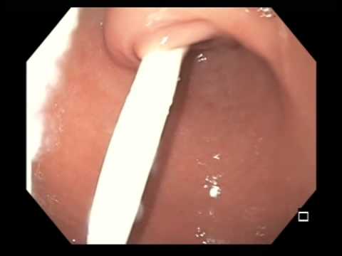
This patient had vomiting of non-bilious fluid following a gastrostomy tube replacement. She was not vomiting tube feeds. The balloon of the replacement tube was within the bulb of the duodenum. Video donated to the SAGES Video Atlas of Endoscopy by Eric M Pauli, MD Keyword(s): balloon, bulb, duodenum, gastric outlet obstruction, non-bilious fluid, post-pyloric […]
Coffee Grounds after Upper GI Bleeding
This video demonstrates one stigmata of a recent upper GI bleed, the finding of ‘coffee ground’ fluid within the stomach. This patient presented with hematemesis and had a small mallory weiss tear not shown on this video. Video donated to the SAGES Video Atlas of Endoscopy by Eric M Pauli, MD Keyword(s): coffee grounds, fluid, […]
Vertical Banded Gastroplasty
This video demonstrates the normal gastric anatomy following a vertical banded gastroplasty. Video donated to the SAGES Video Atlas of Endoscopy by Eric M Pauli, MD Keyword(s): gastric anatomy, VBG
Stomach, Abnormal Post Surgical Anatomy, Gastric Banc with Full Anterior Wall Erosion
This video demonstrates a gastric band that has eroded through the anterior gastric wall. This is well demonstrated in this retroflexed view of the dark staining of the band material due to exposure to gastric contents. Video donated to the SAGES Video Atlas of Endoscopy by Eric M Pauli, MD, Ann M. Rogers, MD Keyword(s): […]
Pyloro-duodenal Stent
This video shows the gastric view of a stent placed across a pyloric stricture. Video donated to the SAGES Video Atlas of Endoscopy by Eric M Pauli, MD Keyword(s): gastric view, pyloric stricture, pyloro-duodenal stent
Hill Grade III/Grade IV Gastroesophageal Valve
This video demonstrates a Hill Grade III/Grade IV gastroesophageal valve. The is opening around esophagus is not tight, and the squamous epithelium can be viewed from below. This represents a very small (Type I) hiatal hernia. Video donated to the SAGES Video Atlas of Endoscopy by Eric M Pauli, MD Keyword(s): esophagus, Hill grade III/IV […]
Gastric Outlet Obestruction from Pyloric Ulcers
This video shows the pylorus in a patient with gastric outlet obstruction. There is edema from ulcers which has lead to partial obstruction of the stomach. Video donated to the SAGES Video Atlas of Endoscopy by Eric M Pauli, MD Keyword(s): gastric outlet obstruction, pyloric ulcers
Cameron’s Ulcer and Hiatal Hernia
This video shows a small cameron’s ulcer associated with a type I Hiatal hernia. There is ulceration on the mucosal folds at the diaphragmatic impression. Video donated to the SAGES Video Atlas of Endoscopy by Eric M Pauli, MD Keyword(s): Camerons ulcer, diaphragmatic impression, mucosal folds, type I hiatal hernia, ulceration
Type III Hiatal Hernia – 2
This video shows a Type III hiatal hernia containing the majority of the patients stomach. The scope loops several times as it is used to reduce the stomach from the chest. Video donated to the SAGES Video Atlas of Endoscopy by Eric M Pauli, MD Keyword(s): chest, stomach, type III hiatal hernia
Stomach, Abnormal Post Surgical Anatomy, Dilated Sleeve Gastrectomy with Retained Fundus
This video shows the endoscopic view post sleeve gastrectomy. The gastric sleeve appears dilated with a capacious fundic component. The retroflexed view of the retained fundus demonstrates this well. Video donated to the SAGES Video Atlas of Endoscopy by Eric M Pauli, MD Keyword(s): capacious fundic component, endoscopic view, fundus, post surgical anatomy, retroflexed view, […]