Normal Appendiceal Orifice, Cecum and Ileocecal Valve
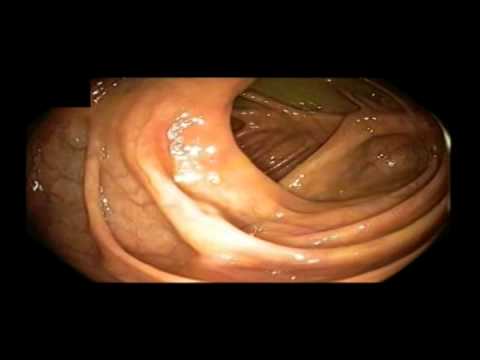
This video demonstrates the normal view of the cecal pit, the appendiceal orifice and the ileocecal valve. Video donated to the SAGES Video Atlas of Endoscopy by Eric M Pauli, MD Keyword(s): appendiceal orifice, cecal pit, cecum, ileocecal valve
Appendiceal Orifice

This viceo shows the colonoscopic view of a normal appendiceal oriface at the base of the cecum. Video donated to the SAGES Video Atlas of Endoscopy by Eric M Pauli, MD Keyword(s): appendiceal orifice, cecum, colonoscopic view
Benign Stricture – Obstruction of Colorectal Anastomosis
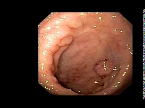
This video demonstrates a near complete obstruction due to a benign anastomotic stricture of a colorectal anastomosis. The ball of granulation tissue on the right of the screen is covering a 2 mm opening that represents the residual anastomosis. Donated to the SAGES Video Atlas of Endoscopy by Eric M Pauli, MD Keyword(s): benign stricture, […]
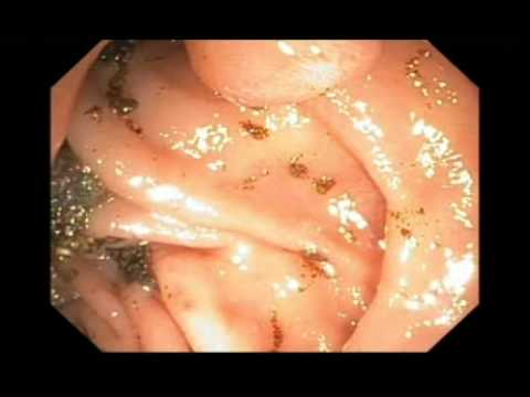
Cecal Sessile Serrated Adenoma

This video demonstrates a sessile, serrated adenoma of the right colon. Video donated to the SAGES Video Atlas of Endoscopy by Eric M Pauli, MD Keyword(s): Cecal Sessile Serrated Adenoma, right colon
Cecal Tubular Adenoma Adjacent to the Ileocecal Valve
This video demonstrates a tubular adenoma of the cecum found adjacent to the ileocecal valve. An endoscopic snare is used to display the full view of the polyp as well as show its proximity to the valve. Video donated to the SAGES Video Atlas of Endoscopy by Eric M Pauli, MD Keyword(s): cecal tubular adenoma, […]