• Skip to primary navigation
  • Skip to content
  • Skip to primary sidebar
  • Skip to footer

SAGES

Society of American Gastrointestinal and Endoscopic Surgeons

  • Home
    • Search
    • SAGES Home
    • SAGES Foundation Home
  • About
    • Committees
      • Descriptions and Video Updates
      • Request to Join a SAGES Committee
      • SAGES Board of Governors
      • Officers and Representatives of the Society
      • Committee Chairs and Mentors
      • Full Committee Rosters
      • SAGES Past Presidents
    • Donate to the SAGES Foundation
    • SAGES Mission Statement
    • SAGES Resource Guide
    • Awards
      • George Berci Award
      • Pioneer in Surgical Endoscopy
      • Excellence In Clinical Care
      • International Ambassador
      • IRCAD Visiting Fellowship
      • Distinguished Service
      • Young Researcher
      • Researcher in Training
      • Jeff Ponsky Master Educator
      • Excellence in Medical Leadership
      • The Brandeis
      • Advocacy Summit Award
      • RAFT Annual Meeting Abstract Contest and Awards
  • Meetings
    • SAGES 2019 Meeting Information
    • 2019 Scientific Session Call For Abstracts
    • 2019 Emerging Technology Session Call For Abstracts
    • 2018 Prevent BDI Consensus Conference
    • CME Claim Form
    • Industry
      • Advertising Opportunities
      • Exhibit Opportunities
      • Sponsorship Opportunities
    • Future Meetings
    • Past Meetings
      • Leadership Development and Health Policy Conference Videos
      • SAGES Quality Summit Meeting
      • SAGES 2018
      • SAGES 2017
      • SAGES 2016
      • SAGES 2015
      • SAGES 2014
      • SAGES 2013
    • Related Meetings Calendar
  • Membership
    • Join SAGES!
    • Give the Gift of SAGES Membership
    • Membership Benefits
    • Active Membership
    • Allied Health Membership
    • Associate Active Membership
    • Candidate Membership
    • International Membership
    • Medical Student Membership
  • For Patients
    • Healthy Sooner – Patient Information for Minimally Invasive Surgery
    • Patient Information Brochures
    • Find a SAGES Member
  • Publications
    • SAGES Clinical / Practice / Training Guidelines, Statements, and Standards of Practice
    • Patient Information Brochures
    • TAVAC – Technology and Value Assessments
    • Surgical Endoscopy and Other Journal Information
    • SAGES Manuals
    • SCOPE – The SAGES Newsletter
    • Troubleshooting Guides
  • Education
    • The Safe Cholecystectomy Didactic Modules
    • Masters Series
      • SAGES Masters Program Facebook Collaboratives
      • Acute Care Surgery
      • Bariatric
      • Biliary
      • Colorectal
      • Flexible Endoscopy (upper or lower)
      • Foregut
      • Hernia
      • Robotics
    • Educational Opportunities
    • The SAGES HPB/Solid Organ Program
    • The SAGES Safe Cholecystectomy Program
    • Courses for Residents
      • Advanced Courses
      • Basic Courses
    • Endorsed Courses
    • SAGES Robotics Fellows Course
    • MIS Fellows Course
    • Free Educational Webinars
    • SMART Enhanced Recovery Program
    • SAGES Quality Initiative
    • SAGES OR SAFETY Video
    • SAGES.TV Video Library
    • SAGES Surgical WIKI
    • iMAGES Image Library
    • SAGES at Cine-Med
      • SAGES Top 21 MIS Procedures
      • SAGES Pearls
      • SAGES Flexible Endoscopy 101
      • SAGES Tips & Tricks of the Top 21
  • Opportunities
    • SAGES Research Opportunities
    • SAGES Endorsed Courses
    • Fundamentals of Laparoscopic Surgery
    • Fundamentals of Endoscopic Surgery
    • Fundamental Use of Surgical Energy
    • Job Board
    • SAGES Go Global: Global Affairs and Humanitarian Efforts
  • Search
    • Search All SAGES Content
    • Search the Video Library
  • Log In

3D structure reconstruction of lesions in colon with colonoscopic images

Atul Kumar1, Shih-Wei Huang, Dr2, Yen-Yu Wang1, Kai-Che Liu1, Ching-Chun Huang1, Yi-Chun Lee1, Jing-Jim Ou2, Wan-Chi Hung1. 1Chang Bing Show Chwan Memorial Hospital, IRCAD-Taiwan, 2Chang Bing Show Chwan Memorial Hospital

Objective: Conventional colonoscopy can only provide a 2D image of a lesion; however, a 3D detailed morphology of the lesions would be helpful to understand the type of lesion and its extension.  This study aims towards developing a system that would help in visualizing a dense 3D structure of colon segment and lesion associated with it.

Description of the technology and method of its use (Fig 1 to 4): A shape from shading (SfS) algorithm is applied to each frame of the colonoscopic video to generate a 3D shape from the image. The algorithm first calculates the direction of the illumination in the image and then finds the depth for each pixel coordinate in the image by solving a non-linear equation. Characteristic feature points in the first frame (F1  ) of the video sequence and the subsequent frame (Fn ) are detected using the speeded-up robust feature (SURF) detection algorithm. The detected interest points are assigned feature point descriptor known as binary string feature point descriptor (BRIEF) which is based upon the intensity differences among the pixels near the interest points. For matching the descriptors, a nearest neighbor search is used with Hamming distance measurements in the descriptor space.  RANdom Sample Consensus (RANSAC) is applied to remove the outliers. The pixel positions of the points of correspondence (between two consecutive images calculated by the feature matching) provide their 2D coordinates (X, Y). Their Z co-ordinates are retrieved from their respective 3D surfaces from SfS. A rigid body transformation matrix between these matched points is calculated using a scaled ICP (Iterative Closest Point) algorithm. The matrix is used to register the 3D surfaces from consecutive video frames. Hence, all the 3D surfaces are stitched together. The pixel coordinates in 2D image and its corresponding depth calculated with SfS provided the x, y and z point coordinates of each pixel in the image. A Delaunay triangulation technique is used to make a 3D surface from the point coordinates. The points on the surface are mapped and interpolated with the color of the respective pixel in 2D image. The final 3D surface is visualized in a 3D render window.

Preliminary results: The complete method was applied using 100 frames of a colonoscopic video from a phantom.   A 3D structure of the lesion and adjoining colon was reconstructed using the method and visualized in a render window. The results is shown in Fig.4.

Conclusions / future directions: The current study presents a method for reconstructing 3D structure of lesions from colonoscopic images. SfS method does not provide a metric structure; therefore, the future study would include information from structure from motion to create a metric scaled structure. The major challenge in the method is to identify characteristic feature points in the colonoscopic images. In future works, feature detection method would be improved further.

Endoscope image of phantom

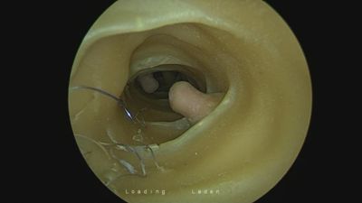
Fig 1. Endoscope image of phantom

Depth map

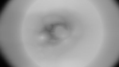
Fig 2. Depth map of endoscope image


Fig 3: Matched features in the endoscope image frames


3D structure from colonoscopic images


Fig 4: 3D structure from the colonoscopic images.

Post Views: 59

Share this:

  • Twitter
  • Facebook
  • LinkedIn
  • Pinterest
  • WhatsApp
  • Reddit

Related

« Return to SAGES 2016 abstract archive

Recently, on SAGES…

Robotic Surgery

Spotlight: Robotics Committee

In the most recent issue of SCOPE, SAGES President Dr. Jeffrey Marks shared his desire to highlight the hard work of the many SAGES committees. As the Society strives to fully realize the new SAGES University Masters Program, the initial focus will be on the affiliated pathway committees and task forces. This is the third […]

Mastering the Art of Surgery – Annual Meeting Press Release

Mastering the Art of Surgery At the 16th World Congress of Endoscopic Surgery and 2018 SAGES Meeting April 11th–14th in Seattle, WA The Society of American Gastrointestinal and Endoscopic Surgeons (SAGES) will kick off its 2018 Annual Meeting as part of the 16th World Congress of Endoscopic Surgery. Hosted by SAGES and CAGS (Canadian Association […]

Complete the SAGES Video Based Assessment Survey for a Chance to Win!

https://survey.az1.qualtrics.com/jfe/form/SV_5pCM9JWvDBphkjj Who Can Enter Open to any surgeon who is either a member of SAGES or an attendee at the 2018 SAGES Annual Meeting AND who completes the 2018 SAGES Video Based Assessment Online Survey at the link above. Drawing Entry Period The drawing period is from survey launch on April 6, 2018 to 11:59pm […]

Contact SAGES

Society of American Gastrointestinal and Endoscopic Surgeons
11300 W. Olympic Blvd Suite 600
Los Angeles, CA 90064 USA
webmaster@sages.org
Tel: (310) 437-0544

Find Us Around the Web!

  • Facebook
  • Twitter
  • YouTube

Newsletter Subscription

  • This field is for validation purposes and should be left unchanged.

Important Links

SAGES 2019 Meeting Information

Online Registration coming in Fall 2018

Healthy Sooner: Patient Information

  • taTME Study Info
  • Foundation
  • SAGES.TV
  • MyCME
  • Educational Activities

· Log in

Copyright © 2019 Society of American Gastrointestinal and Endoscopic Surgeons · Legal
· Managed by BSC Management, Inc