Caroline G Cao, PhD, Peter Y Wong, PhD, Jessica Eisenstein, MS. Tufts University, Medford MA
INTRODUCTION
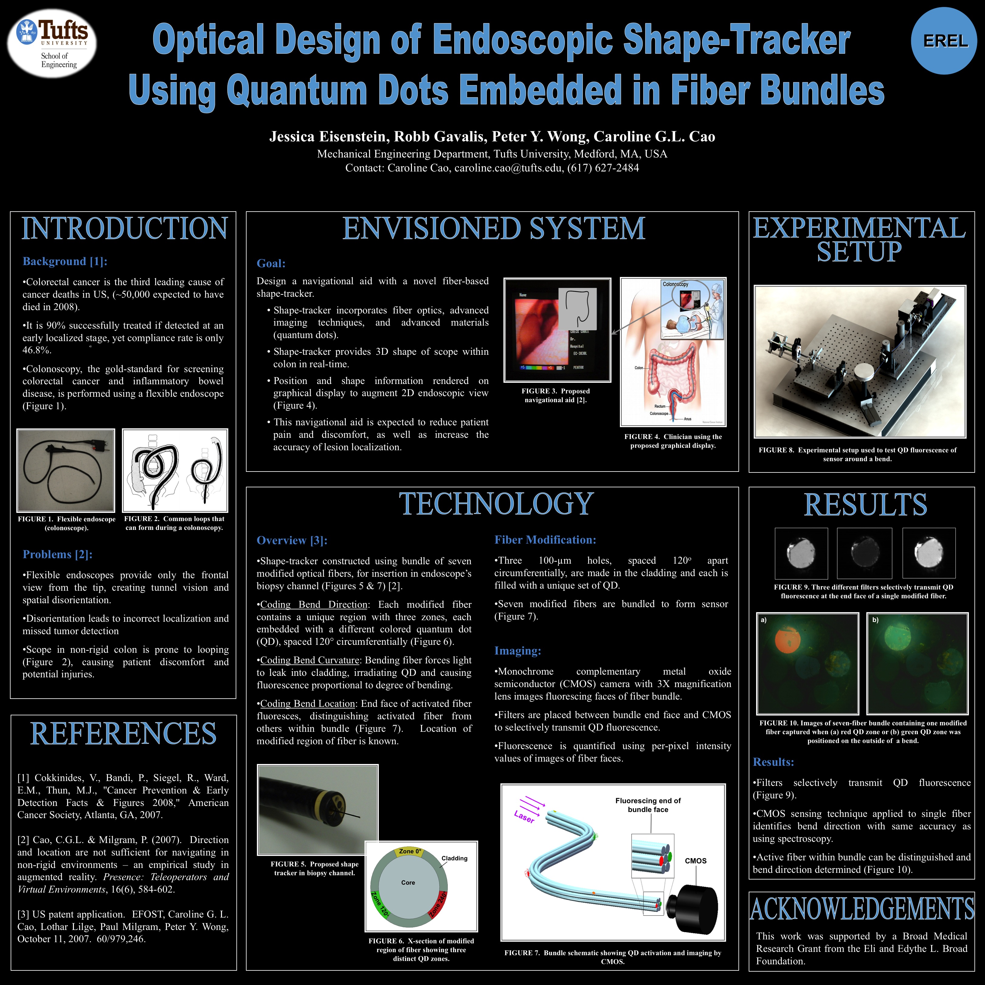
Colon cancer is estimated to be the third leading cause of cancer-related death in the US, with the cost of colorectal cancer treatment reaching $8.4 billion annually. Though colonoscopy is the current standard for colon cancer screening and diagnosis, the procedure has disadvantages due to the flexible scope and the near-blind navigation process used. Loops, which result from the flexible nature of the colon, occur in 91% of cases and are a contributor to disorientation. Disorientation can lead to missed detection of lesions and incorrect localization of tumors.
Research has shown that providing additional spatial information, such as shape, location, and direction of the scope, in a visual display reduces localization errors during colonoscopy. The navigational aid, presented in an augmented reality display, can be superimposed on the standard colonoscopy display. To create the navigational aid, it is necessary to track the shape and orientation of the endoscope in real-time. We describe here a novel imaging technique for the shape-tracker, which uses fiber optics and quantum dots (QDs).
METHODS
Three optical fibers (300 μm silica core, 30 μm polymer cladding), each containing a sensorized region, were bundled together to form a shape-tracking sensor. Three laser-cut QD-modified zones, spaced 120° circumferentially around the fiber, comprised the sensorized region. A dichroic mirror coupled 50mW of 404nm laser light into the bundle. During bending, light leaked from fibers’ cores due to bend loss entered the QD zones positioned on the outer portion of the bend. A complementary metal-oxide-semiconductor (CMOS) camera at the sensor’s proximal end detected the fluorescence from the activated QD to indicate bending direction, curvature, and location.
Evaluation of the shape-tracker was performed inside a colonoscope. Fluorescence data captured by CMOS from these tests were correlated to the direction and degree of bend. The bending information was used to render a graphical image showing the scope’s path and shape. This navigational aid was displayed on a television monitor alongside the endoscopic image for user testing. Eleven different bending direction and curvature combinations were created in a simulated colon environment. Ten novice subjects used the navigational aid to determine the changing shape of the colonoscope during intubation of simulated colons.
RESULTS AND DISCUSSION
Preliminary results demonstrated that CMOS detection of bend loss-induced fluorescence is an effective means of distinguishing bending direction, curvature, and location. Direction and degree of curvature were correctly identified in 100% and 87% of the 15 trials performed, respectively.
FUTURE WORK
Future efforts on the sensor will include increasing the number of fibers within the bundle to thirty-one to characterize the full length of a 1.8m colonoscope, as well as reducing reflections and improving calibration to enhance sensor accuracy. Verification of sensor functionality within the colonoscope will also be performed.
Session: Emerging Technology Poster
Program Number: ETP062
View Poster