Rotating wheels on the headpiece of the endoscope

Rotating wheels on the headpiece of the endoscope control tip deflection. Instruments may be passed through an access port, which is kept capped when not in use (to prevent loss of insufflation and splashing of fluids).
Creating myotomy with a laparoscopic myotome

A. The myotomy is created with a laparoscopic myotome. Make this incision relatively shallow. Traction is applied by a grasper through the righthand port.
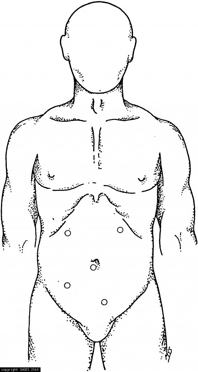
Demonstration of port placement

Demonstration of port placement for repair of a ventral hernia in the upper abdomen. Place the first trocar in the lower midline, 2 or 3 in. inferior to the ventral hernia. Ventral hernias in the lower abdomen require placement of the camera port in the u
Trocar Placement
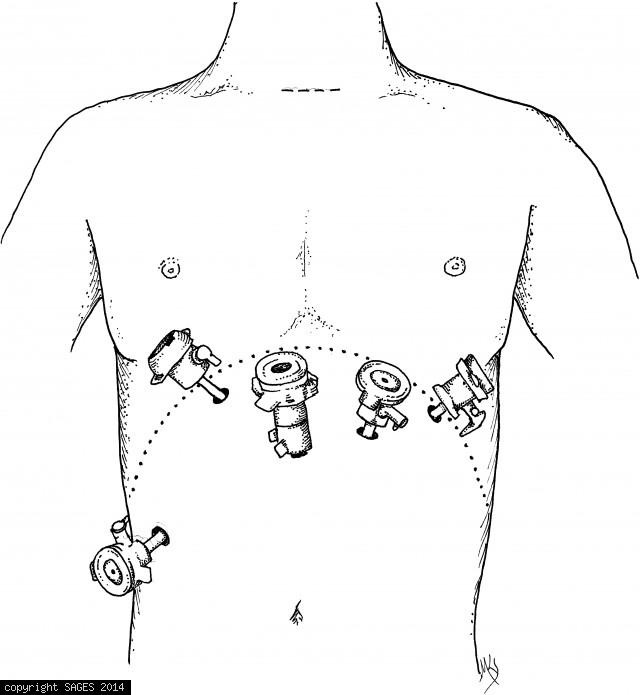
Trocar placement for transabdominal right adrenalectomy. Four 10-mm working trocars are used; if a vascular stapler is needed, one of these can be changed to a 12-mm port.
Trocar Placement
Placement of trocars in laparoscopic transabdominal left adrenalectomy. Three 10-mm trocars are usually placed initially; if a vascular stapler is needed, the middle trocar can be changed to a 12-mm port.
Laparoscopic Splenectomy
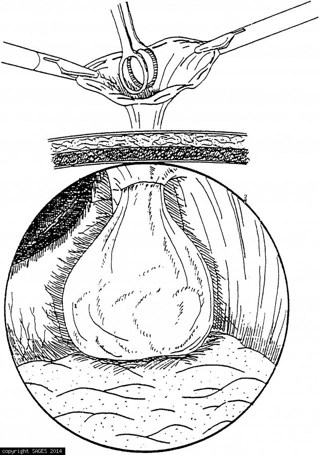
The plastic specimen bag has been retrieved through a large port site. A ring forceps is used to fragment the spleen and remove it piecemeal.

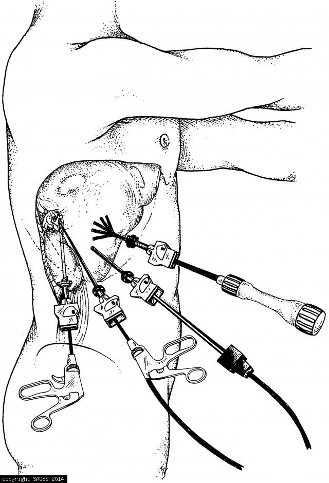
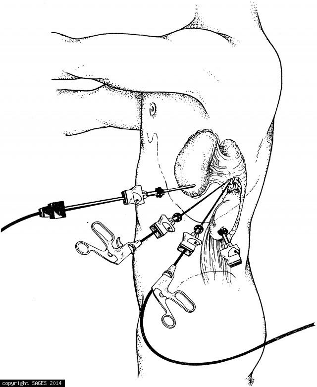
Extracorporeal knot
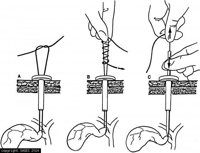
Extracorporeal knot. Bring a long suture into the laparoscopic field, leaving its tail outside the port. Place the stitch; then bring the needle end out through the same port. Create a Roeder knot by tying an overhand knot and then wrapping the suture tai