Colonoscopic Polypectomy
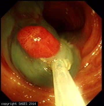
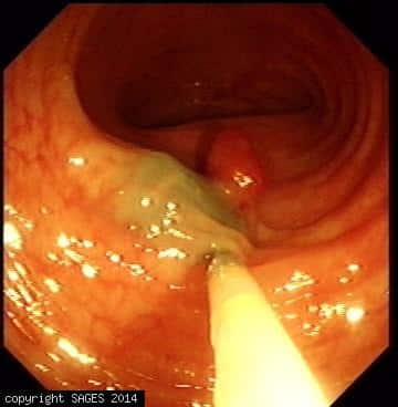
Surgical field — Colonoscopic view. This is the Colonoscopic view of a sessile sigmoid colon adenomatous polyp. Ingection of Methylene blue stained saline beneath the tumor to rise the mucosa from sub mucosa to facilitate safe snare resection.
Colonoscopic Polypectomy
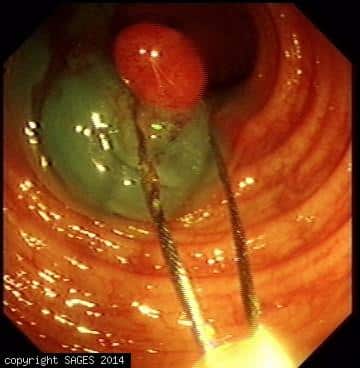
Surgical field — Colonoscopic view. This is the Colonoscopic view of a sessile sigmoid colon adenomatous polyp. Ingection of Methylene blue stained saline beneath the tumor to rise the mucosa from sub mucosa to facilitate safe snare resection.
Colonoscopic Polypectomy
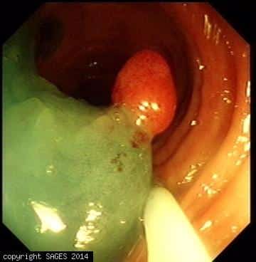
Surgical field — Colonoscopic view. This is the Colonoscopic view of a sessile sigmoid colon adenomatous polyp. Ingection of Methylene blue stained saline beneath the tumor to rise the mucosa from sub mucosa to facilitate safe snare resection.
Colonoscopic Polypectomy
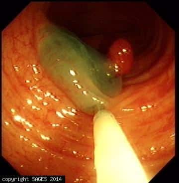
Surgical field — Colonoscopic view. This is the Colonoscopic view of a sessile sigmoid colon adenomatous polyp. Ingection of Methylene blue stained saline beneath the tumor to rise the mucosa from sub mucosa to facilitate safe snare resection.
Colonoscopic Polypectomy
Surgical field — Colonoscopic view. This is the Colonoscopic view of a sessile sigmoid colon adenomatous polyp. Ingection of Methylene blue stained saline beneath the tumor to rise the mucosa from sub mucosa to facilitate safe snare resection.