• Skip to primary navigation
  • Skip to main content
  • Skip to primary sidebar

SAGES

Reimagining surgical care for a healthier world

  • Home
    • Search
    • SAGES Home
    • SAGES Foundation Home
  • About
    • Awards
    • Who Is SAGES?
    • Leadership
    • Our Mission
    • Advocacy
    • Committees
      • SAGES Board of Governors
      • Officers and Representatives of the Society
      • Committee Chairs and Co-Chairs
      • Committee Rosters
      • SAGES Past Presidents
  • Meetings
    • SAGES NBT Innovation Weekend
    • SAGES Annual Meeting
      • 2026 Scientific Session Call for Abstracts
      • 2026 Emerging Technology Call for Abstracts
    • CME Claim Form
    • SAGES Past, Present, Future, and Related Meeting Information
    • SAGES Related Meetings & Events Calendar
  • Join SAGES!
    • Membership Application
    • Membership Benefits
    • Membership Types
      • Requirements and Applications for Active Membership in SAGES
      • Requirements and Applications for Affiliate Membership in SAGES
      • Requirements and Applications for Associate Active Membership in SAGES
      • Requirements and Applications for Candidate Membership in SAGES
      • Requirements and Applications for International Membership in SAGES
      • Requirements for Medical Student Membership
    • Member Spotlight
    • Give the Gift of SAGES Membership
  • Patients
    • Join the SAGES Patient Partner Network (PPN)
    • Patient Information Brochures
    • Healthy Sooner – Patient Information for Minimally Invasive Surgery
    • Choosing Wisely – An Initiative of the ABIM Foundation
    • All in the Recovery: Colorectal Cancer Alliance
    • Find A SAGES Surgeon
  • Publications
    • Sustainability in Surgical Practice
    • SAGES Stories Podcast
    • SAGES Clinical / Practice / Training Guidelines, Statements, and Standards of Practice
    • Patient Information Brochures
    • Patient Information From SAGES
    • TAVAC – Technology and Value Assessments
    • Surgical Endoscopy and Other Journal Information
    • SAGES Manuals
    • MesSAGES – The SAGES Newsletter
    • COVID-19 Archive
    • Troubleshooting Guides
  • Education
    • Wellness Resources – You Are Not Alone
    • Avoid Opiates After Surgery
    • SAGES Subscription Catalog
    • SAGES TV: Home of SAGES Surgical Videos
    • The SAGES Safe Cholecystectomy Program
    • Masters Program
    • Resident and Fellow Opportunities
      • SAGES Free Resident Webinar Series
      • Fluorescence-Guided Surgery Course for Fellows
      • Fellows’ Career Development Course
      • SAGES Robotics Residents and Fellows Courses
      • MIS Fellows Course
    • SAGES S.M.A.R.T. Enhanced Recovery Program
    • SAGES @ Cine-Med Products
      • SAGES Top 21 Minimally Invasive Procedures Every Practicing Surgeon Should Know
      • SAGES Pearls Step-by-Step
      • SAGES Flexible Endoscopy 101
    • SAGES OR SAFETY Video Activity
  • Opportunities
    • Fellowship Recognition Opportunities
    • SAGES Advanced Flexible Endoscopy Area of Concentrated Training (ACT) SEAL
    • Multi-Society Foregut Fellowship Certification
    • Research Opportunities
    • FLS
    • FES
    • FUSE
    • Jobs Board
    • SAGES Go Global: Global Affairs and Humanitarian Efforts
  • Search
    • Search the SAGES Site
    • Guidelines Search
    • Video Search
    • Search Images
    • Search Abstracts
  • OWLS/FLS
  • Login
You are here: Home / Archives for suture

Image Tag: Suture

Braided suture snared by the endoscopist

The braided suture is snared by the endoscopist and will be drawn back through the esophagus to exit through the mouth.

Posted on
06/16/2010
Image Categories
Fundamentals: Percutaneous Endoscopic, SAGES Manual
Image Tags
braided endoscopist, esophagus, mouth, snared, suture

Method of mesh fixation

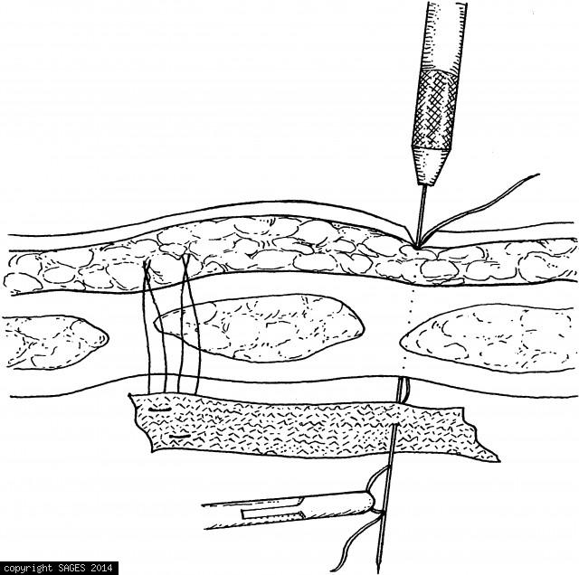
Method of mesh fixation with a suture passer. Use the suture passer to introduce the suture through the musculofascial layer, the mesh, and then back out through all layers as a mattress suture. Use four to eight mattress sutures to anchor the mesh, depen

Posted on
06/15/2010
Image Categories
Fundamentals: Repair of Ventral Hernia, SAGES Manual
Image Tags
abdominal wall, anchor, hernia, knot, mattress, mesh, musculofascial, staples, subcutaneously, suture

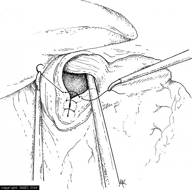
Firing and Removing the Stapler

The stapler has been fired and removed. The staple line has been inspected for hemostasis and is being closed with a simple running suture.

Posted on
06/15/2010
Image Categories
Fundamentals:Cholecystojejunostomy&Gastro, SAGES Manual
Image Tags
hemostasis, stapler, suture

Inspecting the staple line for hemostasis

After the staple line has been inspected for hemostasis, the enterotomies are closed with a running suture. (Reprinted with permission from Bogen GL, Mancino AT, Scott-Conner CE. Laparoscopy for staging and palliation of gastrointestinal malignancy. Surg

Posted on
06/15/2010
Image Categories
Fundamentals:Cholecystojejunostomy&Gastro, SAGES Manual
Image Tags
enterotomies, gastrointestinal, hemostasis, malignancy, palliation, staple, suture

Trocar Placement

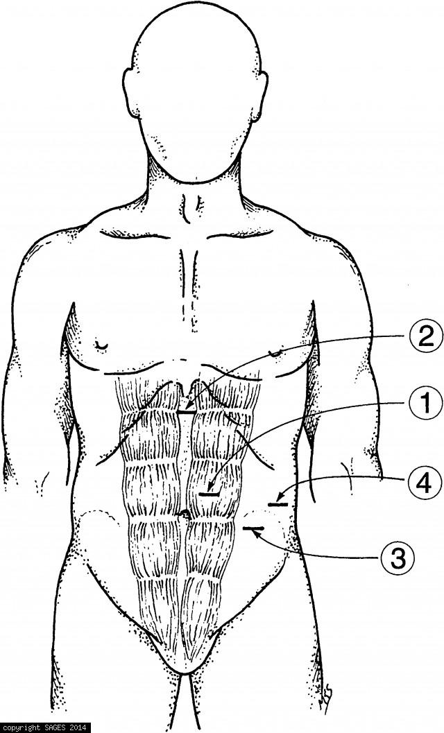
Trocar placements for distal pancreatectomy. These sites are proper for both splenic salvage and with splenectomy. There should be at least a hand’s breadth distance between trocars 1, 3, and 4. The trocar for the laparoscope should be above the umbilicus

Posted on
06/15/2010
Image Categories
Fundamentals: Distal Pancreatectomy, SAGES Manual
Image Tags
anastomosis, distal, gastric, gastrointestinal, knot tying, pancreatectomy, splenectomy, suture, trocar, vessels

The anvil of the circular stapler

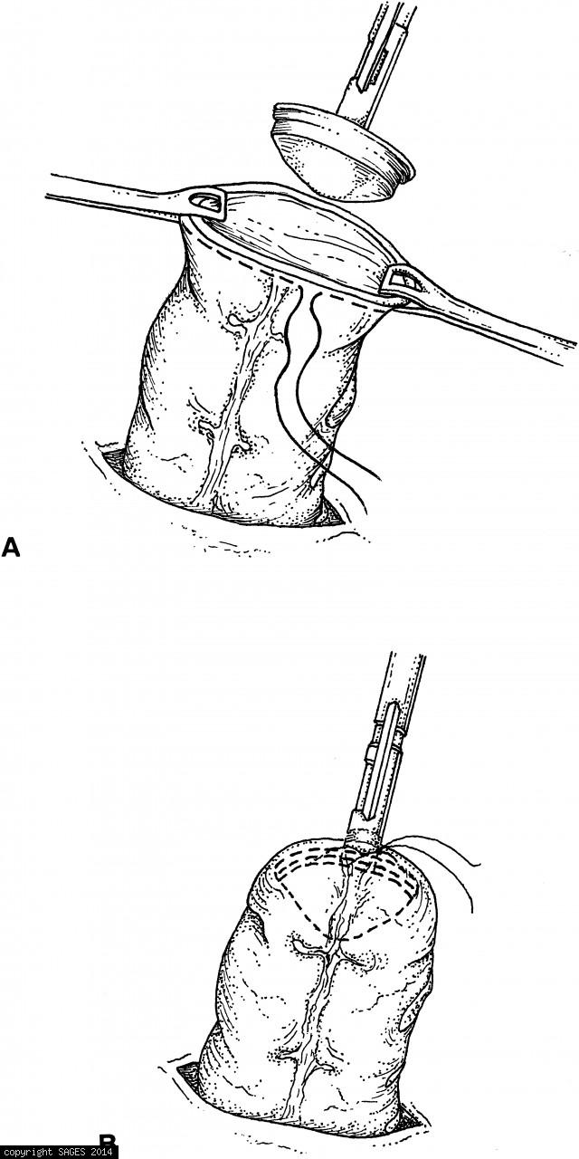
A. The anvil of the circular stapler is inserted in the proximal end of the bowel (which has been drawn out of the abdomen through an enlarged trocar site). B. The pursestring suture is tied. The bowel is then returned to the abdomen.

Posted on
06/14/2010
Image Categories
Fundamentals: Segmental Colectomies, Anterior, SAGES Manual
Image Tags
abdomen, anvil, stapler, suture

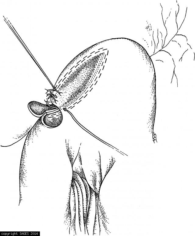
Closure of the Crural Opening

The crural opening is closed with simple, interrupted nonabsorbable suture (0 Ethibond). For large defects, pledgets and/or bioprosthetic mesh may be used.

Posted on
06/11/2010
Image Categories
Fundamentals: Paraesophageal Hernia Repair, SAGES Manual
Image Tags
bioprosthetic mesh, crucal opening, pledgets, suture

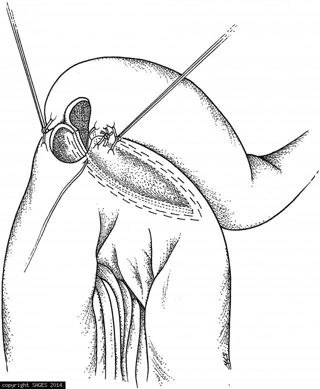
Proper Orientation of the Fundic Wrap

The fundoplication is sutured in place with a single U-stitch of 2–0 Prolene pledgeted on the outside. A 60-French mercury-weighted bougie is passed through the gastroesophageal junction prior to fixation of the wrap to assure a floppy fundoplication. Ins

Posted on
06/11/2010
Image Categories
Fundamentals: Treatment of Gastroesophageal, SAGES Manual
Image Tags
fundoplication, gastroesophageal junction, suture

Extracorporeal knot

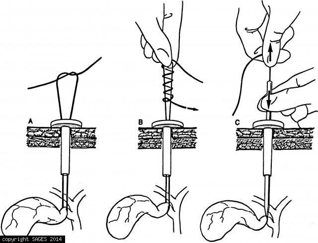
Extracorporeal knot. Bring a long suture into the laparoscopic field, leaving its tail outside the port. Place the stitch; then bring the needle end out through the same port. Create a Roeder knot by tying an overhand knot and then wrapping the suture tai

Posted on
06/10/2010
Image Categories
Fundamentals: Principles of Tissue Approxim., SAGES Manual
Image Tags
extracorporeal knot, needle, port, suture

Search and Filters

Search by keyword(s)

Filter by image tag(s)

Filter by image category(s)

  • Foundation
  • SAGES.TV
  • MyCME
  • Educational Activities

Copyright © 2025 Society of American Gastrointestinal and Endoscopic Surgeons