• Skip to primary navigation
  • Skip to main content
  • Skip to primary sidebar

SAGES

Reimagining surgical care for a healthier world

  • Home
    • Search
    • SAGES Home
    • SAGES Foundation Home
  • About
    • Awards
    • Who Is SAGES?
    • Leadership
    • Our Mission
    • Advocacy
    • Committees
      • SAGES Board of Governors
      • Officers and Representatives of the Society
      • Committee Chairs and Co-Chairs
      • Committee Rosters
      • SAGES Past Presidents
  • Meetings
    • SAGES NBT Innovation Weekend
    • SAGES Annual Meeting
      • 2026 Scientific Session Call for Abstracts
      • 2026 Emerging Technology Call for Abstracts
    • CME Claim Form
    • SAGES Past, Present, Future, and Related Meeting Information
    • SAGES Related Meetings & Events Calendar
  • Join SAGES!
    • Membership Application
    • Membership Benefits
    • Membership Types
      • Requirements and Applications for Active Membership in SAGES
      • Requirements and Applications for Affiliate Membership in SAGES
      • Requirements and Applications for Associate Active Membership in SAGES
      • Requirements and Applications for Candidate Membership in SAGES
      • Requirements and Applications for International Membership in SAGES
      • Requirements for Medical Student Membership
    • Member Spotlight
    • Give the Gift of SAGES Membership
  • Patients
    • Join the SAGES Patient Partner Network (PPN)
    • Patient Information Brochures
    • Healthy Sooner – Patient Information for Minimally Invasive Surgery
    • Choosing Wisely – An Initiative of the ABIM Foundation
    • All in the Recovery: Colorectal Cancer Alliance
    • Find A SAGES Surgeon
  • Publications
    • Sustainability in Surgical Practice
    • SAGES Stories Podcast
    • SAGES Clinical / Practice / Training Guidelines, Statements, and Standards of Practice
    • Patient Information Brochures
    • Patient Information From SAGES
    • TAVAC – Technology and Value Assessments
    • Surgical Endoscopy and Other Journal Information
    • SAGES Manuals
    • MesSAGES – The SAGES Newsletter
    • COVID-19 Archive
    • Troubleshooting Guides
  • Education
    • Wellness Resources – You Are Not Alone
    • Avoid Opiates After Surgery
    • SAGES Subscription Catalog
    • SAGES TV: Home of SAGES Surgical Videos
    • The SAGES Safe Cholecystectomy Program
    • Masters Program
    • Resident and Fellow Opportunities
      • SAGES Free Resident Webinar Series
      • Fluorescence-Guided Surgery Course for Fellows
      • Fellows’ Career Development Course
      • SAGES Robotics Residents and Fellows Courses
      • MIS Fellows Course
    • SAGES S.M.A.R.T. Enhanced Recovery Program
    • SAGES @ Cine-Med Products
      • SAGES Top 21 Minimally Invasive Procedures Every Practicing Surgeon Should Know
      • SAGES Pearls Step-by-Step
      • SAGES Flexible Endoscopy 101
    • SAGES OR SAFETY Video Activity
  • Opportunities
    • Fellowship Recognition Opportunities
    • SAGES Advanced Flexible Endoscopy Area of Concentrated Training (ACT) SEAL
    • Multi-Society Foregut Fellowship Certification
    • Research Opportunities
    • FLS
    • FES
    • FUSE
    • Jobs Board
    • SAGES Go Global: Global Affairs and Humanitarian Efforts
  • Search
    • Search the SAGES Site
    • Guidelines Search
    • Video Search
    • Search Images
    • Search Abstracts
  • OWLS/FLS
  • Login
You are here: Home / Archives for stomach

Image Tag: Stomach

View of Pylorus

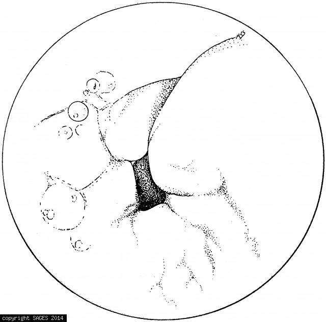
View of pylorus. Bubbles indicating bile provide a clue to the location of the distal stomach when orientation of the side-viewing endoscope is difficult.

Posted on
06/16/2010
Image Categories
Fundamentals: Endoscopic Retrograde, SAGES Manual
Image Tags
bile, bubbles, distal, endoscope, pylorus, stomach

PEG tube

The PEG tube is pulled back into the stomach. Endoscopic verification of placement is essential.

Posted on
06/16/2010
Image Categories
Fundamentals: Percutaneous Endoscopic, SAGES Manual
Image Tags
endoscopic, peg tube, stomach

Transillumination and finger depression

A. Transillumination and finger depression of the abdominal wall confirm juxtaposition of the inflated stomach and the anterior abdominal wall. B. The site selected will generally be approximately halfway between costal margin and umbilicus.
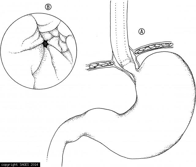
Posted on
06/16/2010
Image Categories
Fundamentals: Percutaneous Endoscopic, SAGES Manual
Image Tags
abdominal wall, anterior, costal margin, finger depression, inflated, juxtaposition, stomach, transillumination, umbilicus

Patient Positioning

With the patient in the left lateral decubitus position, the endoscopist facing the patient, and the scope relaxed as described in the text, entry into the stomach will generally give a view oriented with the lesser curvature at 12 o’clock, the greater cu

Posted on
06/16/2010
Image Categories
Fundamentals: Diagnostic Upper, SAGES Manual
Image Tags
anterior, curvature, decubitus, endoscopist, lateral, patient, posterior, stomach

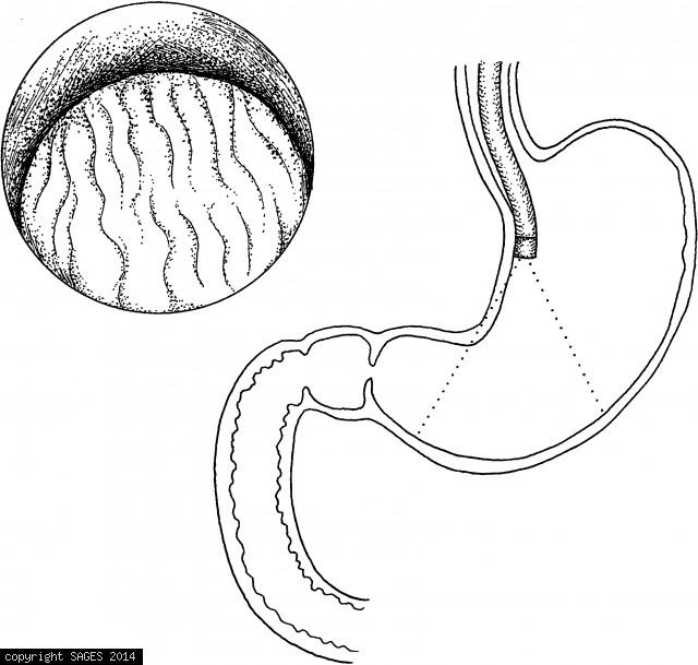
Upper Gastrointestinal Endoscopy

A. The endoscope is advanced down the relatively straight esophagus until the lower esophageal sphincter is identified. B. The lower esophageal sphincter often coincides with the transition from squamous epithelium (white) of the esophagus to mucosa (pink

Posted on
06/16/2010
Image Categories
Fundamentals: Diagnostic Upper, SAGES Manual
Image Tags
endoscope, epithelium, esophagus, mucosa, sphincter, squamous, stomach

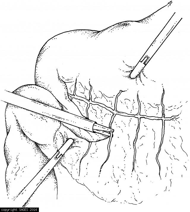
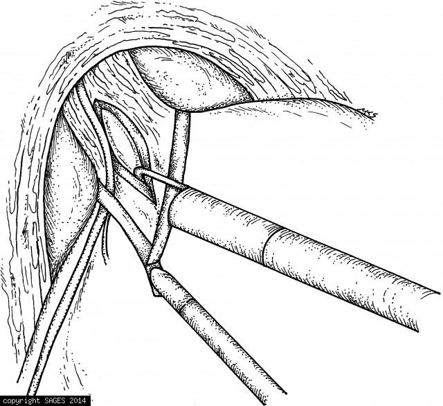
Selecting and Affixing loop of jejunum

A loop of jejunum has been selected and affixed to the greater curvature of the stomach, above the gastroepiploic vessels, with two stay sutures. Two enterotomies have been made and the stapling device inserted.

Posted on
06/15/2010
Image Categories
Fundamentals:Cholecystojejunostomy&Gastro, SAGES Manual
Image Tags
enterotomies, gastroepiploic, jejunum, loop, stomach, sutures, vessels

Trocar Placement

Trocar placement for gastrojejunostomy is slightly different, in that trocar 2 is placed lower, to allow adequate working distance from the stomach. If you plan to do both procedures, use this trocar arrangement (rather than that in Fig. 36.1).

Posted on
06/15/2010
Image Categories
Fundamentals:Cholecystojejunostomy&Gastro, SAGES Manual
Image Tags
gastrojejunostomy, stomach, trocar

Retracting the stomach cephalad

The stomach is retracted cephalad as the transverse colon is retracted caudad while the omentum is divided with the harmonic scalpel.

Posted on
06/14/2010
Image Categories
Fundamentals: Assisted Proctocolectomy, SAGES Manual
Image Tags
caudad, cephalad, harmonic, omentum, retracted, scalpel, stomach, transverse colon

Gastric Balloon catheter to facilitate pouch

The stomach is exposed and mobilized. A gastric balloon catheter may be used to define the pouch and facilitate pouch creation. (Soper NJ, Swanstrom LL, Eubanks WS, eds. Mastery of Endoscopic and Laparoscopic Surgery. 2d ed. Philadelphia: Lippincott Willi

Posted on
06/14/2010
Image Categories
Fundamentals: Bariatric Srgy. Patient Select., SAGES Manual
Image Tags
gastric balloon, pouch creation, stomach

Sling retraction of the stomach

Sling retraction of the stomach.

Posted on
06/11/2010
Image Categories
Fundamentals: Gastric Resections, SAGES Manual
Image Tags
sling retraction, stomach

Insertion of the Innerdyne gastric cannula

Insertion of the Innerdyne gastric cannula into inflated stomach. Thereafter, the radially expandable polymer sheath is stretched by an 11.0-mm port for intragastric insertion of the operating laparoscope.

Posted on
06/11/2010
Image Categories
Fundamentals: Gastric Resections, SAGES Manual
Image Tags
innerdyne gastric cannula, intragastric insertion, polymer sheath, stomach

Construction of mucosa-lined tube

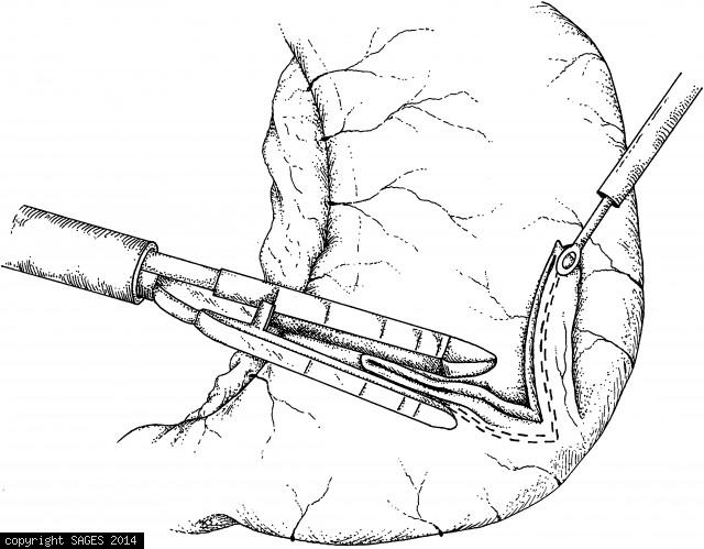
Construction of mucosa-lined tube (Janeway-style gastrostomy). A fold of stomach is elevated and the endoscopic stapler applied. Approximately 1 cm of stomach must be included in the staple line to assure an adequate lumen. The tube is grasped and elevate

Posted on
06/11/2010
Image Categories
Fundamentals: Laparoscopic Gastrostomy, SAGES Manual
Image Tags
cannula, janewaystyle, lumen, mucosa, stapler, stomach

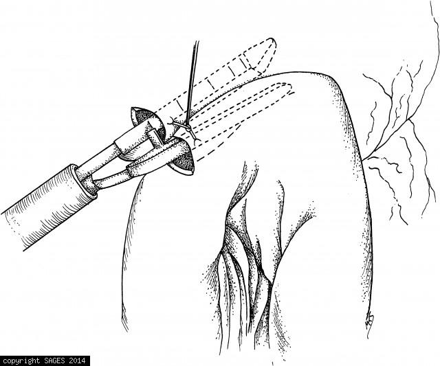
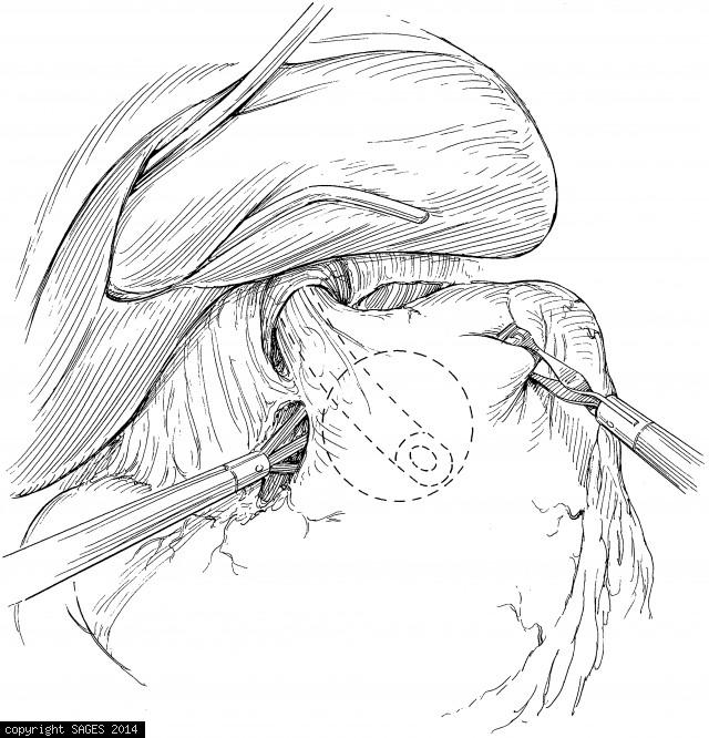
Myotomy carried distally by hook Electrocautery

The myotomy being carried distally, using hook electrocautery. Care must be taken to elevate the muscle fibers away from the mucosa before the electrocautery is applied. The myotomy extends about 5 to 6 cm proximal to the gastroesophageal junction and abo

Posted on
06/11/2010
Image Categories
Fundamentals: Cardiomyotomy (Heller Myotomy), SAGES Manual
Image Tags
electrocautery, gastroesophageal junction, mucosa, muscle fibers, myotomy, stomach

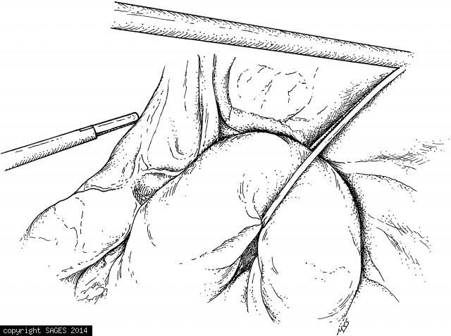
Mobilization of the fundus

Retract the stomach rightward and the spleen and omentum left and downward to complete mobilization of the fundus. These maneuvers open the lesser sac and facilitate division of the high short gastric vessels.

Posted on
06/11/2010
Image Categories
Fundamentals: Treatment of Gastroesophageal, SAGES Manual
Image Tags
gastric vessels, mobilization of the fundus, spleen, stomach

Dissection of the left crus

Dissection of the left crus. The left crus is dissected as completely as possible, and the attachments of the fundus of the stomach to the diaphragm are taken down.

Posted on
06/11/2010
Image Categories
Fundamentals: Treatment of Gastroesophageal, SAGES Manual
Image Tags
cruss, diaphram, dissection, fundus, stomach

Search and Filters

Search by keyword(s)

Filter by image tag(s)

Filter by image category(s)

  • Foundation
  • SAGES.TV
  • MyCME
  • Educational Activities

Copyright © 2025 Society of American Gastrointestinal and Endoscopic Surgeons