Meckels diverticulum
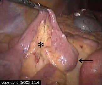
Meckels found during diagnostic laparoscopy for unknown GI bleeding.Surgical field—Laparoscopic view of Meckel`s diverticula. Mesodiverticula (asterisk); Tip of the diverticulum containing heterotopic tissue (arrow).
Video monitor and Cart
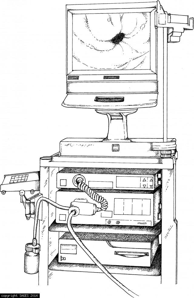
Cart with video monitor, light source, video processor, water bottle, and image printer. A keyboard allows entry of patient and physician names, patient number, date, and any additional documentation desired.
Trocar site placement colon resections

Trocar site placement for laparoscopic pull-through and other colon resections.

Trocar site placement for undescended testicle

Trocar site placement for surgery for undescended testicle.

Hypertrophied Segment division
C. This division is continued through the hypertrophied segment, but not onto the duodenum.
Pyloromyotomy spreader to divide hypertorphied

B. A pyloromyotomy spreader is used to divide the hypertrophied circular muscle fibers.
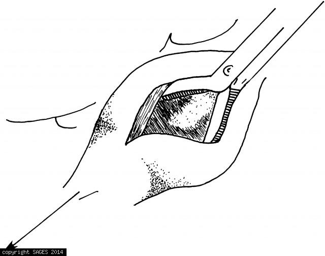
Creating myotomy with a laparoscopic myotome

A. The myotomy is created with a laparoscopic myotome. Make this incision relatively shallow. Traction is applied by a grasper through the righthand port.
Patient Position and Trocar Sites
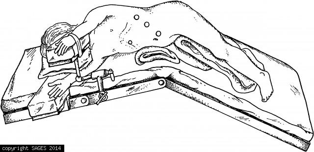
Patient position and trocar sites for pediatric laparoscopic splenectomy. Note that the patient is in the lateral position, and the operating table has been flexed to increase the distance between costal margin and superior iliac crest.
Trocar placement
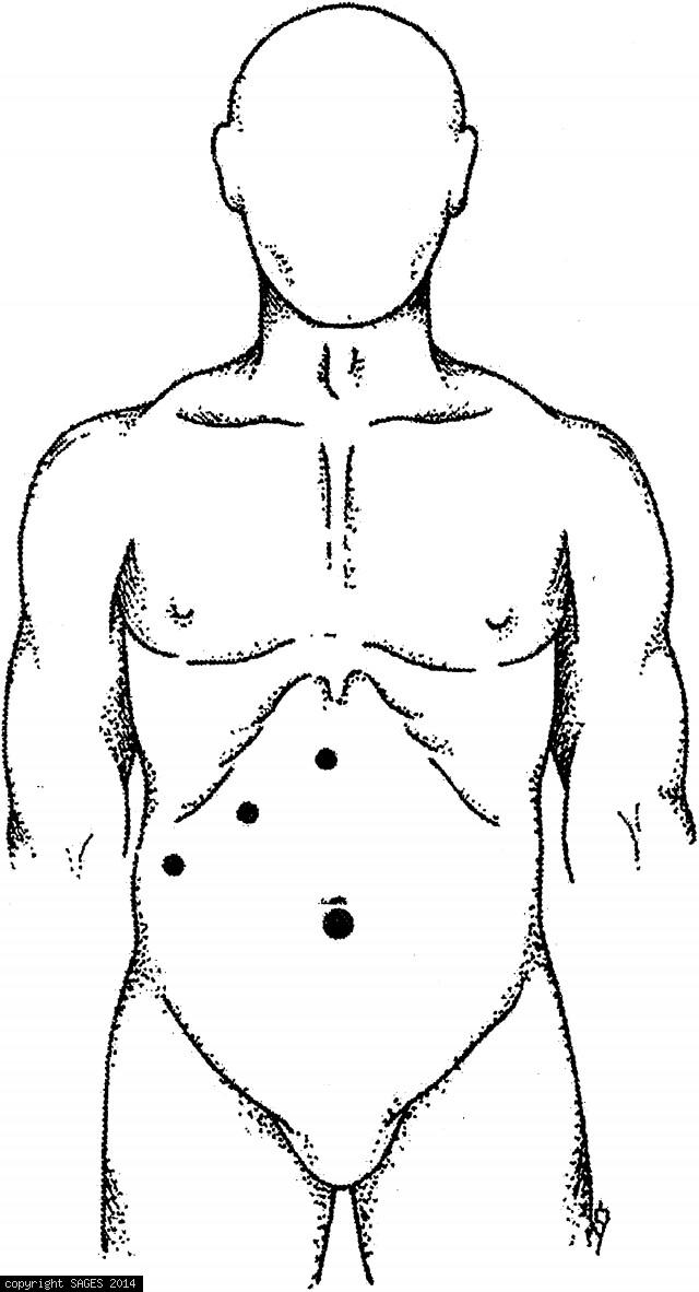
Trocar site placement for pediatric laparoscopic cholecystectomy is similar to the adult procedure.
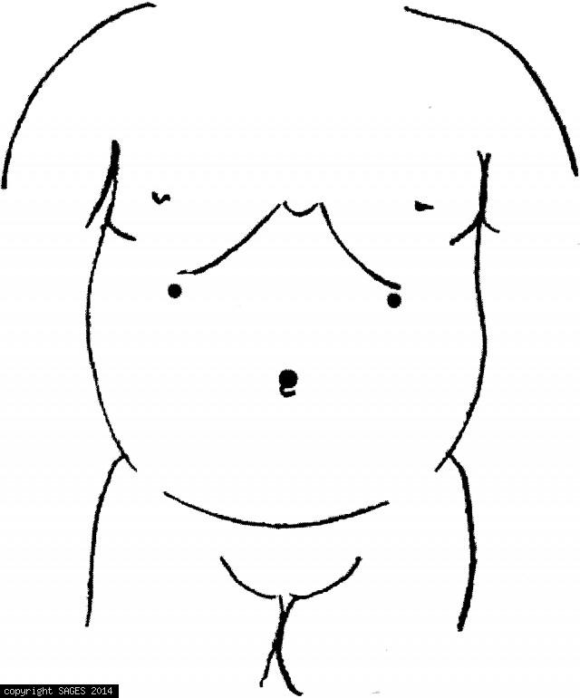
Trocar sites for laparoscopic appendectomy
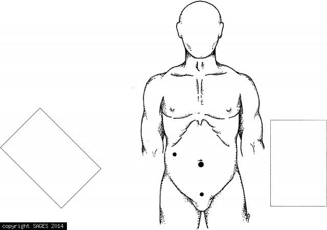
Trocar sites for laparoscopic appendectomy (alternate placement) with two monitors. Surgeon stands to the left, assistant may need to stand to the left.