GIST
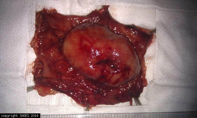
A case of gastric gastrointestinal stromal tumor (GIST), underwent laparoscopic wide local excision with free margin.
Huge large Sliding Hiatal Hernia

Example of hudge sliding hiatal(esophageal) hernia. On the right side, this is forward and reflexed endoscopic view (Retrovision maneuver)in a patient demonstrating huge sliding hiatal hernia. On the left side, the laparoscopic view of the same patient.
Standardization of Video-assisted Esophagectomy: Hand-assisted Thoracoscopic Surgery As a Safety Procedure

Authors: Toshiaki Shichinohe, MD PhD, Shunichi Okushiba, MD PhD, You Kawarada, MD PhD, Shuji Kitashiro, MD PhD, Hiroto Manase, MD PhD, Kentaro Kato, MD PhD, Takahiro Tsuchikawa, MD PhD, Joe Matsumoto, MD PhD, Ryosuke Kawasaki, MD PhD, Eiichi Tanaka, MD P
Lower Levels of Occasional Blood Glucose in Patients with Billroth I Reconstruction after Not Vagus Nerve-spared Laparoscoy-assisted Distal Gastrectom
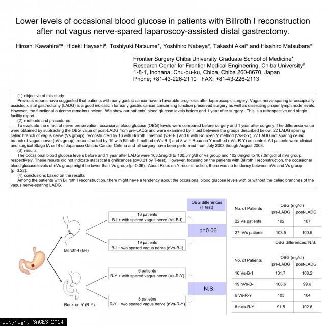
Authors: Hiroshi Kawahira, MD, Hideki Hayashi, MD, Toshiyuki Natsume, MD, Yoshihiro Nabeya, MD, Takashi Akai, MD, Hisahiro Matsubara, MD
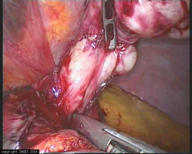
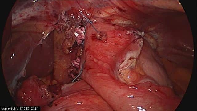
Gastroesophageal Junction GIST tumor
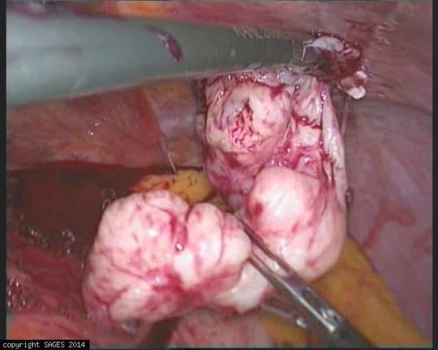
Example of Gastroesophageal GIST tumor on Laparoscopy.This is the laparoscopic view in a patient demonstrating big Gastroesophageal GIST presenting with nausea and vomiting, treated by laparoscopic resection.
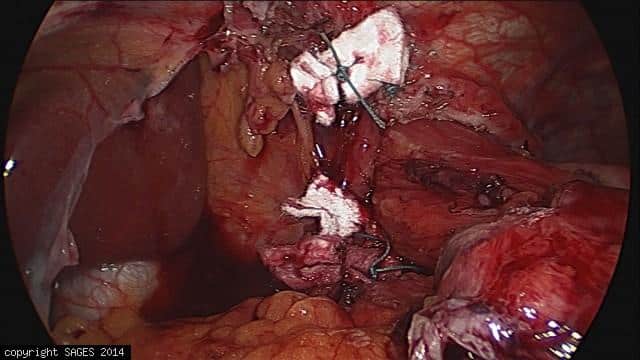
Gastroesophageal Junction GIST tumor
Example of Gastroesophageal GIST tumor on Laparoscopy.This is the laparoscopic view in a patient demonstrating big Gastroesophageal GIST presenting with nausea and vomiting, treated by laparoscopic resection.
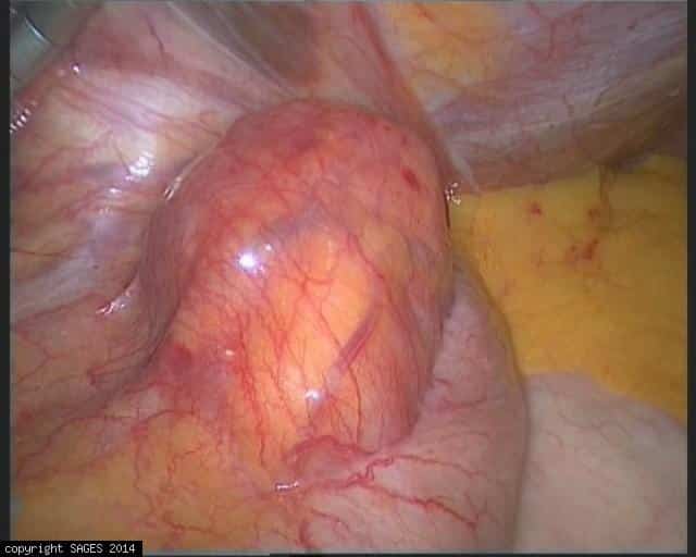
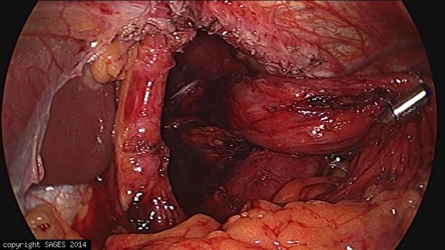
Gastroesophageal Junction GIST tumor

Example of Gastroesophageal GIST tumor on Laparoscopy.This is the laparoscopic view in a patient demonstrating big Gastroesophageal GIST presenting with nausea and vomiting, treated by laparoscopic resection.
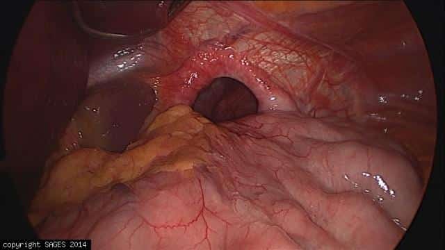
Gastroesophageal Junction GIST tumor
Example of Gastroesophageal GIST tumor on Laparoscopy.This is the laparoscopic view in a patient demonstrating big Gastroesophageal GIST presenting with nausea and vomiting, treated by laparoscopic resection.
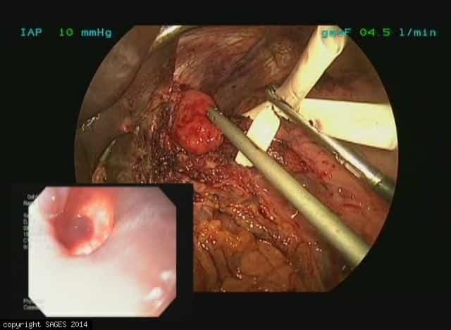
Gastroesophageal Junction GIST tumor

Gastroesophageal GIST tumor on Endoscopy.This is the forward and reflex view (Retrovision maneuver)in a patient demonstrating big Gastroesophageal GIST presenting with nausea and vomiting, treated by laparoscopic resection(images:102151-102154)
Laparoscopic Revision of Esophagomyotomy
A 53 year old man with several months history of dysphagia and apreistaltic esophagus had undergone a laparoscopic esophagomyotomy in early 2008 with short lived symptom relief. The operative report indicated the myotomy had been carried well up on the e
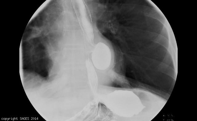
UGI of Slipped Nissen
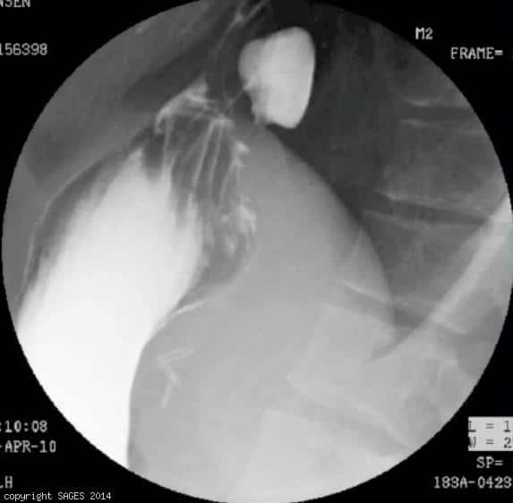
35 year old female with history of Nissen, now with dysphagia and GERD, UGI shows partial slippage into the chest.
Paraesophageal Hernia – anterior fundoplication
This is an image of an anterior fundoplication after repair of a paraesophageal hernia
Paraesophageal Hernia – repaired
This is an image of a paraesophageal hernia after primary suture repair
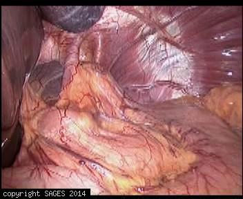
Paraesophageal Hernia – dissected
This is an image of the hiatus with a paraesophageal hernia after dissection
Paraesophageal Hernia – reduced
This is an image of a paraesophageal hernia after reduction of the entire stomach from the chest
Paraesophageal hernia before reduction

This is an image of the esophageal hiatus with a paraesophageal hernia with the entire stomcah in the chest
Epiphrenic Diverticulum
66 year-old male with epiphrenic diverticulum and diffuse esophageal spasm dysmotility.
New Concept of Hybrid Esophagectomy to Enhance Its Safety and Radicality for Esophageal Cancer
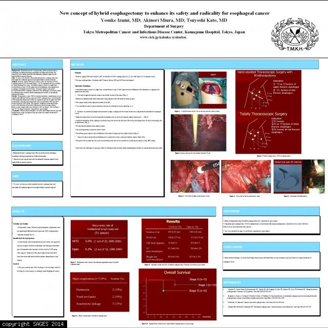
Authors: Yosuke Izumi, MD, Akinori Miura, MD, Tsuyoshi Kato, MD
Longitudinal and circular fibers during Heller myotomy

This photo show the disdection during Heller myotomy. The left shows both circular and longitudinal fibers visualized and the right show this section of the myotomy completed