Endoscopic visualization of the ampulla of Vater
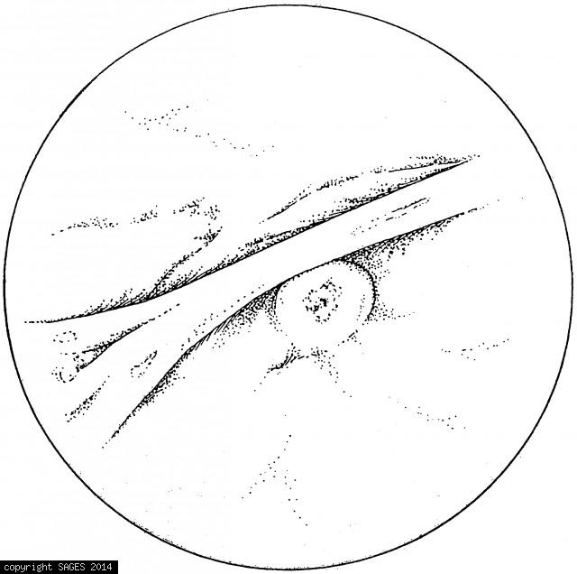
Endoscopic visualization of the ampulla of Vater. A transverse fold of mucosa overlying the ampulla is frequently seen, as shown here.
Upper Gastrointestinal Endoscopy
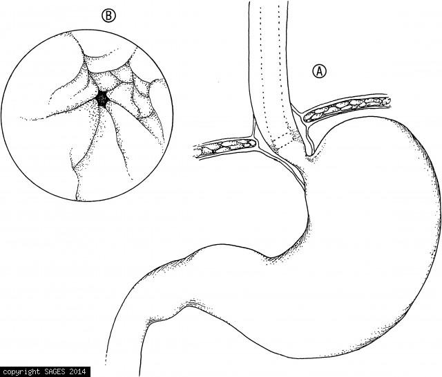
A. The endoscope is advanced down the relatively straight esophagus until the lower esophageal sphincter is identified. B. The lower esophageal sphincter often coincides with the transition from squamous epithelium (white) of the esophagus to mucosa (pink
Construction of mucosa-lined tube
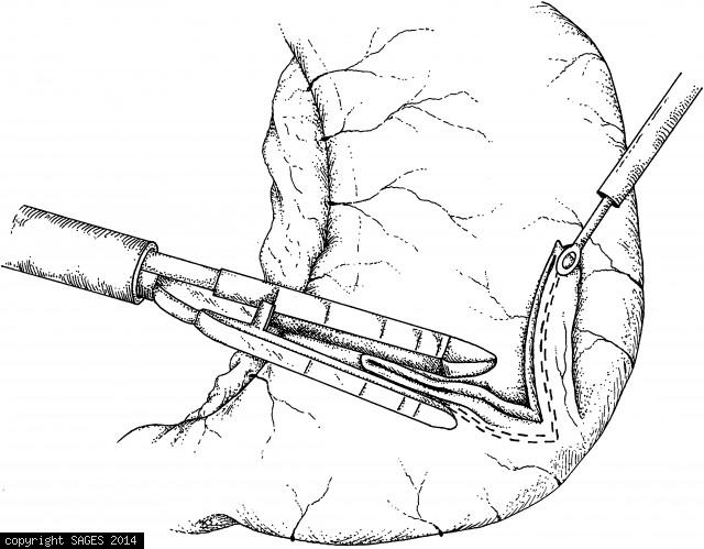
Construction of mucosa-lined tube (Janeway-style gastrostomy). A fold of stomach is elevated and the endoscopic stapler applied. Approximately 1 cm of stomach must be included in the staple line to assure an adequate lumen. The tube is grasped and elevate
Myotomy carried distally by hook Electrocautery
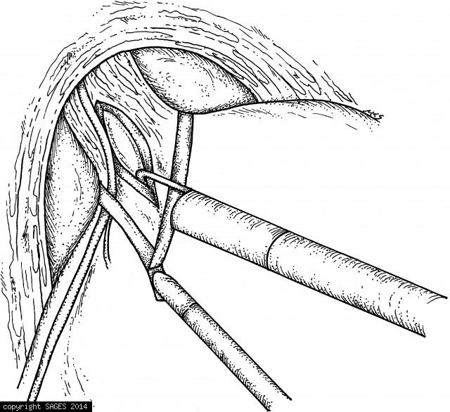
The myotomy being carried distally, using hook electrocautery. Care must be taken to elevate the muscle fibers away from the mucosa before the electrocautery is applied. The myotomy extends about 5 to 6 cm proximal to the gastroesophageal junction and abo