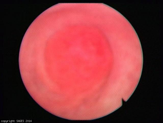
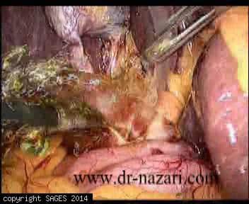
Robot Assisted LCBDE Distal View
Robot assisted laparoscopic common bile duct exploration. Distal view after stone removal.
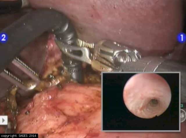
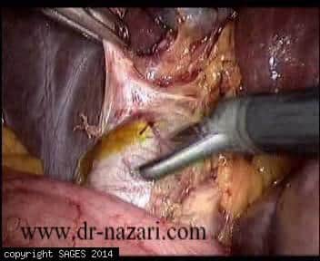
Robot-Assisted LCBDE
Robot assisted laparoscopic common bile duct exploration using choledochoscope.
Intra-Operative Cholangiography
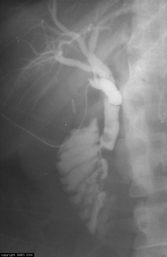
Laparoscopic Intra-Operative cholangiography using catheter inside the cystic duct and instillation of radio-opaque agent showing multiple choledocholithiasis.
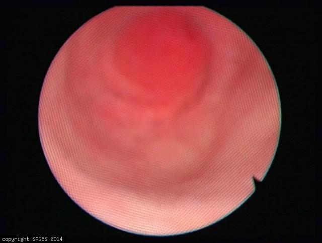
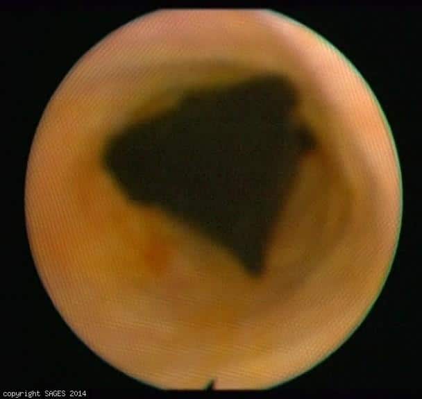
Peripapillary duodenal diverticulum

Choledochoscopic view of peripapillary duodenal diverticulum (type II). Type II of peripapillary duodenal diverticulum makes the elevation of bile duct pressure directly caused by intraduodenal pressure loading. In choledochoscopic view, the diverticulum
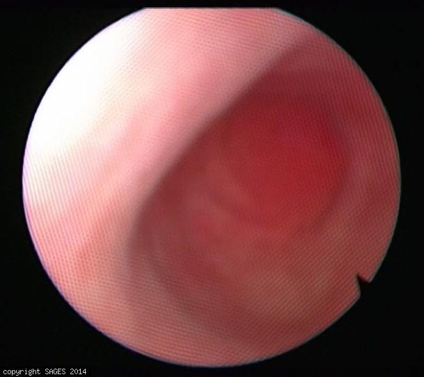
Peripapillary duodenal diverticulum

Choledochoscopic view of peripapillary duodenal diverticulum (type II). Type II of peripapillary duodenal diverticulum makes the elevation of bile duct pressure directly caused by intraduodenal pressure loading. In choledochoscopic view, the diverticulum
Peripapillary duodenal diverticulum

Choledochoscopic view of peripapillary duodenal diverticulum (type II). Type II of peripapillary duodenal diverticulum makes the elevation of bile duct pressure directly caused by intraduodenal pressure loading. In choledochoscopic view, the diverticulum
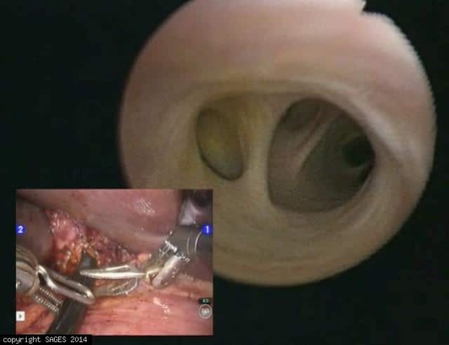
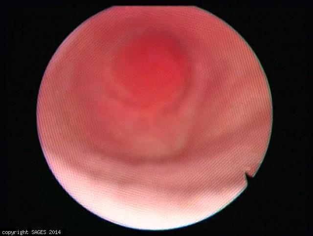
Peripapillary duodenal diverticulum (Inside view)

Choledochoscopic view of peripapillary duodenal diverticulum (type II). Type II of peripapillary duodenal diverticulum makes the elevation of bile duct pressure directly caused by intraduodenal pressure loading. In choledochoscopic view, the diverticulum
Peripapillary duodenal diverticulum

Choledochoscopic view of peripapillary duodenal diverticulum (type II). Type II of peripapillary duodenal diverticulum makes the elevation of bile duct pressure directly caused by intraduodenal pressure loading. In choledochoscopic view, the diverticulum
Peripapillary duodenal diverticulum
Choledochoscopic view of peripapillary duodenal diverticulum (type II). Type II of peripapillary duodenal diverticulum makes the elevation of bile duct pressure directly caused by intraduodenal pressure loading. In choledochoscopic view, the diverticulum
Peripapillary duodenal diverticulum

Choledochoscopic view of peripapillary duodenal diverticulum (type II). Type II of peripapillary duodenal diverticulum makes the elevation of bile duct pressure directly caused by intraduodenal pressure loading. In choledochoscopic view, the diverticulum
Peripapillary duodenal diverticulum

Choledochoscopic view of peripapillary duodenal diverticulum (type II). Type II of peripapillary duodenal diverticulum makes the elevation of bile duct pressure directly caused by intraduodenal pressure loading. In choledochoscopic view, the diverticulum
Peripapillary duodenal diverticulum
Choledochoscopic view of peripapillary duodenal diverticulum (type II). Type II of peripapillary duodenal diverticulum makes the elevation of bile duct pressure directly caused by intraduodenal pressure loading. In choledochoscopic view, the diverticulum
Peripapillary duodenal diverticulum

Choledochoscopic view of peripapillary duodenal diverticulum (type II). Type II of peripapillary duodenal diverticulum makes the elevation of bile duct pressure directly caused by intraduodenal pressure loading. In choledochoscopic view, the diverticulum
Peripapillary duodenal diverticulum (Lap. view)

Choledochoscopic view of peripapillary duodenal diverticulum (type II). Type II of peripapillary duodenal diverticulum makes the elevation of bile duct pressure directly caused by intraduodenal pressure loading. In choledochoscopic view, the diverticulum
Peripapillary duodenal diverticulum

Choledochoscopic view of peripapillary duodenal diverticulum (type II). Type II of peripapillary duodenal diverticulum makes the elevation of bile duct pressure directly caused by intraduodenal pressure loading. In choledochoscopic view, the diverticulum
Peripapillary duodenal diverticulum

Choledochoscopic view of peripapillary duodenal diverticulum (type II). Type II of peripapillary duodenal diverticulum makes the elevation of bile duct pressure directly caused by intraduodenal pressure loading. In choledochoscopic view, the diverticulum
Peripapillary duodenal diverticulum
Choledochoscopic view of peripapillary duodenal diverticulum (type II). Type II of peripapillary duodenal diverticulum makes the elevation of bile duct pressure directly caused by intraduodenal pressure loading. In choledochoscopic view, the diverticulum
Peripapillary duodenal diverticulum
Choledochoscopic view of peripapillary duodenal diverticulum (type II). Type II of peripapillary duodenal diverticulum makes the elevation of bile duct pressure directly caused by intraduodenal pressure loading. In choledochoscopic view, the diverticulum
CBD closure
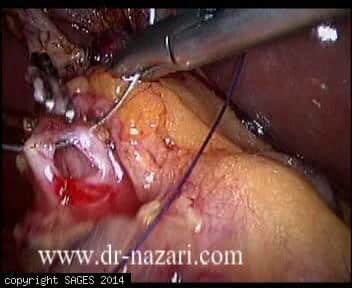
After all stones were retrieved and clearance of the CBD was confirmed with re-choledochoscopy, choledochotomy was closed with interrupted sutures, 3/0 vicryl on a ski needle.
Choledochoscopic basketing of CBD stones.

Common bile duct stones were retrieved using wire (Dormia) basket.
CBD clearance
Clearance of the CBD was attempted by wash out them by instillation of normal saline via a tube inserted in CBD.

Choledochotomy
A longitudinal choledochotomy was made using a Berci knife and microscissors in the supraduodenal part of the CBD.