• Skip to primary navigation
  • Skip to main content

SAGES

Reimagining surgical care for a healthier world

  • Home
    • Search
    • SAGES Home
    • SAGES Foundation Home
  • About
    • Awards
    • Who Is SAGES?
    • Leadership
    • Our Mission
    • Advocacy
    • Committees
      • SAGES Board of Governors
      • Officers and Representatives of the Society
      • Committee Chairs and Co-Chairs
      • Committee Rosters
      • SAGES Past Presidents
  • Meetings
    • SAGES NBT Innovation Weekend
    • SAGES Annual Meeting
      • 2026 Scientific Session Call for Abstracts
      • 2026 Emerging Technology Call for Abstracts
    • CME Claim Form
    • SAGES Past, Present, Future, and Related Meeting Information
    • SAGES Related Meetings & Events Calendar
  • Join SAGES!
    • Membership Application
    • Membership Benefits
    • Membership Types
      • Requirements and Applications for Active Membership in SAGES
      • Requirements and Applications for Affiliate Membership in SAGES
      • Requirements and Applications for Associate Active Membership in SAGES
      • Requirements and Applications for Candidate Membership in SAGES
      • Requirements and Applications for International Membership in SAGES
      • Requirements for Medical Student Membership
    • Member Spotlight
    • Give the Gift of SAGES Membership
  • Patients
    • Join the SAGES Patient Partner Network (PPN)
    • Patient Information Brochures
    • Healthy Sooner – Patient Information for Minimally Invasive Surgery
    • Choosing Wisely – An Initiative of the ABIM Foundation
    • All in the Recovery: Colorectal Cancer Alliance
    • Find A SAGES Surgeon
  • Publications
    • Sustainability in Surgical Practice
    • SAGES Stories Podcast
    • SAGES Clinical / Practice / Training Guidelines, Statements, and Standards of Practice
    • Patient Information Brochures
    • Patient Information From SAGES
    • TAVAC – Technology and Value Assessments
    • Surgical Endoscopy and Other Journal Information
    • SAGES Manuals
    • MesSAGES – The SAGES Newsletter
    • COVID-19 Archive
    • Troubleshooting Guides
  • Education
    • Wellness Resources – You Are Not Alone
    • Avoid Opiates After Surgery
    • SAGES Subscription Catalog
    • SAGES TV: Home of SAGES Surgical Videos
    • The SAGES Safe Cholecystectomy Program
    • Masters Program
    • Resident and Fellow Opportunities
      • SAGES Free Resident Webinar Series
      • Fluorescence-Guided Surgery Course for Fellows
      • Fellows’ Career Development Course
      • SAGES Robotics Residents and Fellows Courses
      • MIS Fellows Course
    • SAGES S.M.A.R.T. Enhanced Recovery Program
    • SAGES @ Cine-Med Products
      • SAGES Top 21 Minimally Invasive Procedures Every Practicing Surgeon Should Know
      • SAGES Pearls Step-by-Step
      • SAGES Flexible Endoscopy 101
    • SAGES OR SAFETY Video Activity
  • Opportunities
    • Fellowship Recognition Opportunities
    • SAGES Advanced Flexible Endoscopy Area of Concentrated Training (ACT) SEAL
    • Multi-Society Foregut Fellowship Certification
    • Research Opportunities
    • FLS
    • FES
    • FUSE
    • Jobs Board
    • SAGES Go Global: Global Affairs and Humanitarian Efforts
  • Search
    • Search the SAGES Site
    • Guidelines Search
    • Video Search
    • Search Images
    • Search Abstracts
  • OWLS/FLS
  • Login
You are here: Home / Abstracts / A method to combine fluorescence image with endoscopic image for undersurface vascular visualization

A method to combine fluorescence image with endoscopic image for undersurface vascular visualization

Shih-Wei Huang, MD1, Atul Kumar2, Hon-Phin Wong, MD1, Hurng-Sheng Wu1, Yen-Yu Wang2, Kai-Che Liu2, Lih-Shun Chen2, Yi-Chun Lee2, Wan-Chi Hung2. 1Show Chwan Memorial Hospital, Changhua, 2Chang Bing Show Chwan Memorial Hospital, Changhua

1. Objective: Identification of undersurface major arteries and veins is a challenge during endoscopic surgery, which is needed to avoid inadvertent injuries. Indocyanine green (ICG) fluorescence has been found to be promising in visualizing the undersurface blood vessels during laparoscopic surgery. However, the fluorescence of the vasculature disappears within few minutes.If a longer visualization of vessels is required, the ICG is injected repeatedly. A system is needed which can preserve the fluorescence information of the vasculature and provide it whenever required during the surgery.This study aims to develop a system that would superimpose the relevant information of fluorescence image from a previous scene on the white light image of the current scene that would be useful for locating relatively static vasculature throughout the surgery.

2 Method
2.1 Video acquisition: Laparoscopic video was acquired with Karl Storz high definition image acquisition system. The vascular area as seen under infra-red camera was simulated by a blue colored drawing of vessels.

2.2 Navigation system

The navigation system consists of a tracking system and a visualization software. An NDI Polaris tracking system was used for this study. The software used the Visualization toolkit and Insight toolkit C++ libraries.

2.3 Segmentation of the vasculature

The region of interest in the registered fluorescence (simulated)  image was segmented using a threshold technique in its blue channel in the RGB color space.A Canny edge detector was applied to calculate the edge of the image.

2.4 Superimposition of the images

The software connected the endoscope camera to the NDI tracking system. An NDI tracking tool was mounted on the camera. The video frame and the respective pose of the camera was recorded. The endoscope image (TargetImage) corresponding to the segmented fluorescence image was stored and visualized as a target image plane with the segmented region of interest. The current frame of the video (CurrentImage) was visualized in the same navigation display. The user moved the camera to match the current image plane (CurrentImage) with the TargetImage when the alignement was needed.

3 Preliminary results

A C ++ based software for the system was being developed in a computer with Intel R CoreTM i7 960@3.20GHz, 6.00RAM 64 bit Windows 7 and graphics card of NVidia TESLA C2075. The software uses the libraries of VTK, OpenCV and ITK. The superimposed view of the white light image and the edge image from the fluorescence (simulated) image  is shown in Figure 2 and 3.

4 Conclusions/future works

This work develops a system which can store the information of the highlighted area in a fluorescence (simulated) image and superimpose the relevant information on a white light whenever needed. In future, the system would be applied on real surgery and the tracking matching would be replaced by a robot arm.

ICG fluorescence image

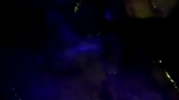
Figure 1. A typical ICG fluorescence image

Edge image and the current image when they are not aligned

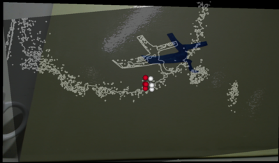
Figure 2: Edge image and current frame not aligned ( The blue region simulates the ICG fluorescence).


Figure 3: Edge image and current frame aligned ( The blue region simulates the ICG fluorescence).


Presented at the SAGES 2017 Annual Meeting in Houston, TX.

Abstract ID: 91083

Program Number: ETP890

Presentation Session: Emerging Technology iPoster Session (Non CME)

Presentation Type: Poster

37


  • Foundation
  • SAGES.TV
  • MyCME
  • Educational Activities

Copyright © 2025 Society of American Gastrointestinal and Endoscopic Surgeons