• Skip to primary navigation
  • Skip to main content

SAGES

Reimagining surgical care for a healthier world

  • Home
    • Search
    • SAGES Home
    • SAGES Foundation Home
  • About
    • Awards
    • Who Is SAGES?
    • Leadership
    • Our Mission
    • Advocacy
    • Committees
      • SAGES Board of Governors
      • Officers and Representatives of the Society
      • Committee Chairs and Co-Chairs
      • Committee Rosters
      • SAGES Past Presidents
  • Meetings
    • SAGES NBT Innovation Weekend
    • SAGES Annual Meeting
      • 2026 Scientific Session Call for Abstracts
      • 2026 Emerging Technology Call for Abstracts
    • CME Claim Form
    • SAGES Past, Present, Future, and Related Meeting Information
    • SAGES Related Meetings & Events Calendar
  • Join SAGES!
    • Membership Application
    • Membership Benefits
    • Membership Types
      • Requirements and Applications for Active Membership in SAGES
      • Requirements and Applications for Affiliate Membership in SAGES
      • Requirements and Applications for Associate Active Membership in SAGES
      • Requirements and Applications for Candidate Membership in SAGES
      • Requirements and Applications for International Membership in SAGES
      • Requirements for Medical Student Membership
    • Member Spotlight
    • Give the Gift of SAGES Membership
  • Patients
    • Join the SAGES Patient Partner Network (PPN)
    • Patient Information Brochures
    • Healthy Sooner – Patient Information for Minimally Invasive Surgery
    • Choosing Wisely – An Initiative of the ABIM Foundation
    • All in the Recovery: Colorectal Cancer Alliance
    • Find A SAGES Surgeon
  • Publications
    • Sustainability in Surgical Practice
    • SAGES Stories Podcast
    • SAGES Clinical / Practice / Training Guidelines, Statements, and Standards of Practice
    • Patient Information Brochures
    • Patient Information From SAGES
    • TAVAC – Technology and Value Assessments
    • Surgical Endoscopy and Other Journal Information
    • SAGES Manuals
    • MesSAGES – The SAGES Newsletter
    • COVID-19 Archive
    • Troubleshooting Guides
  • Education
    • Wellness Resources – You Are Not Alone
    • Avoid Opiates After Surgery
    • SAGES Subscription Catalog
    • SAGES TV: Home of SAGES Surgical Videos
    • The SAGES Safe Cholecystectomy Program
    • Masters Program
    • Resident and Fellow Opportunities
      • SAGES Free Resident Webinar Series
      • Fluorescence-Guided Surgery Course for Fellows
      • Fellows’ Career Development Course
      • SAGES Robotics Residents and Fellows Courses
      • MIS Fellows Course
    • SAGES S.M.A.R.T. Enhanced Recovery Program
    • SAGES @ Cine-Med Products
      • SAGES Top 21 Minimally Invasive Procedures Every Practicing Surgeon Should Know
      • SAGES Pearls Step-by-Step
      • SAGES Flexible Endoscopy 101
    • SAGES OR SAFETY Video Activity
  • Opportunities
    • Fellowship Recognition Opportunities
    • SAGES Advanced Flexible Endoscopy Area of Concentrated Training (ACT) SEAL
    • Multi-Society Foregut Fellowship Certification
    • Research Opportunities
    • FLS
    • FES
    • FUSE
    • Jobs Board
    • SAGES Go Global: Global Affairs and Humanitarian Efforts
  • Search
    • Search the SAGES Site
    • Guidelines Search
    • Video Search
    • Search Images
    • Search Abstracts
  • OWLS/FLS
  • Login
You are here: Home / Abstracts / 3D printing technology for surgical planning models in laparoscopic abdominal surgery

3D printing technology for surgical planning models in laparoscopic abdominal surgery

Stefania Marconi, MSc1, Andrea Pietrabissa, PhD, MD2, Ferdinando Auricchio, PhD3. 1Dept. of Surgical Sciences, Dept. of Civil Engineering and Architecture, University of Pavia, 2Dept. of Surgical Sciences, University of Pavia, 3Dept. of Civil Engineering and Architecture, University of Pavia

Objectives

The aim of the work is the use of 3D printing technology to build patient-specific models for procedure planning in laparoscopic surgery, especially for splenectomies and live donor kidney procurements. Our goal is improving the way surgical procedure are planned, thanks to the informative power of physic objects with respect to conventional imaging systems. The final goal is the integration of these facilities in the daily clinical practice, considering their importance also for educational purposes and as a support for the explanation of the surgical procedure to patients.

Materials and Methods

We start form Multi Detector Computed Tomography (MDCT) image elaboration to virtually reconstruct the patient-specific anatomy: MDCT images are acquired using a contrast dye with a slice-thickness resolution of at least 2 mm. For spleen models only arterial contrastographic phase is used, while for renal models also the late phase is used to reconstruct ureters.

Through the use of an open source software, ITK-Snap (https://www.itksnap.org), we apply a segmentation algorithm that identifies and marks with a specific label the structure of interest in each MDCT slice. Therefore, arteries, veins, ureters and the various parenchymal structures are identified with different labels. The resulting label set can be rendered to get a 3D virtual model of each anatomical structure. Each model is then exported using the STL (Standard Tessellation Language) file format that can be processed for the 3D printing: the STL model is sliced into layers of the same thickness of 3D printer resolution. For our purpose, we use an Objet 30 Pro 3D printer, able to deploy layers up to 16 microns of thickness. The high-resolution is mandatory to reproduce all the fine details of anatomical structures, especially the vascular ones. Models are printed with a white rigid photo-polymeric material, which is deployed in a liquid form and cured with UV light during the printing process.

The image elaboration procedure takes from 1 to 3 hours, depending on image quality, while the printing of each model takes up to 23 hours: after the printing, each structure is painted in different colours and assembled.

Results

Up to now we produced 16 patient-specific 3D printed models: 12 for laparoscopic splenectomy and 4 for live donor kidney procurement. Among splenectomy models, 10 were investigated to assess their ability in transferring detailed information, especially to young surgeons. Moreover, the usefulness of the 3D printed models for the patient comprehension of the surgical procedure has been qualitatively assessed.

Conclusions

3D printed models of patient-specific anatomy can give an important aid to surgical planning, since they can transfer information in a more immediate way: the possibility of hold in hand the model can significantly improve the quality of the planning. Models usefulness is quite evident for young surgeons, since they have less experience in building a mental 3D reconstruction of the specific anatomy. Through this facility, both novice surgeons and patients have the possibility of better understanding the surgical procedure.

image

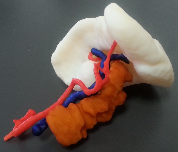
Fig. 1: Example of a 3D printed model for laparoscopic splenectomy.

848


  • Foundation
  • SAGES.TV
  • MyCME
  • Educational Activities

Copyright © 2025 Society of American Gastrointestinal and Endoscopic Surgeons