
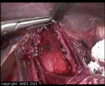
Longitudinal and circular fibers during Heller myotomy

This photo show the disdection during Heller myotomy. The left shows both circular and longitudinal fibers visualized and the right show this section of the myotomy completed
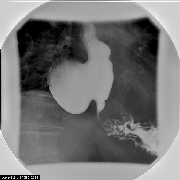
Dilated Esophagus in Achalasia Patient

Coronal views of a dilated esophagus with excessive retained food, secondary to achalasia.
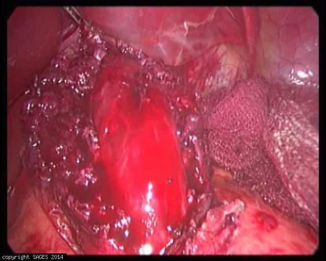
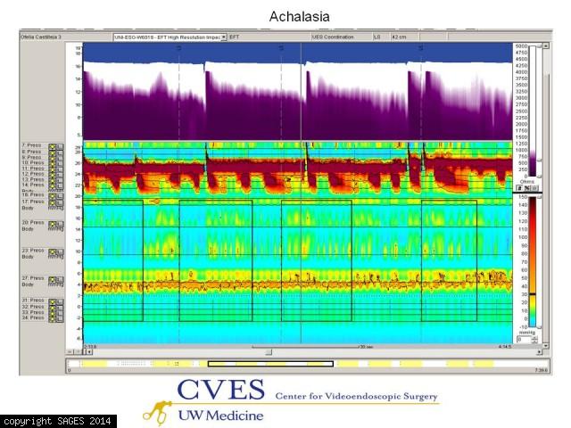
Achalasia
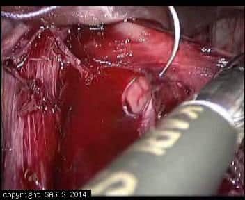
Image obtained during Laparoscopic Heller`s Cardiomyotomy. Intracorporeal suturing of mucosal perforation.
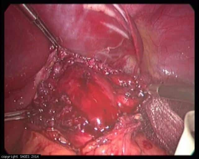
Achalasia
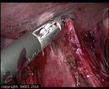
Image obtained during Laparoscopic Heller`s Cardiomyotomy. A mucosal perforation was done during myotomy.


Epiphrenic diverticulum seen on contrast study
Epiphrenic diverticulum seen on barium contrast study in patient with achalasia.