• Skip to main content
  • Skip to header right navigation
  • Skip to site footer

Log in
www.sages.org

SAGES

Reimagining surgical care for a healthier world

  • Home
    • SAGES Home
    • SAGES Foundation Home
  • About
    • Awards
    • Who Is SAGES?
    • Leadership
    • Our Mission
    • Advocacy
    • Committees
      • SAGES Board of Governors
      • Officers and Representatives of the Society
      • Committee Chairs and Co-Chairs
      • Committee Rosters
      • SAGES Past Presidents
    • Why Should You Support SAGES?
    • SAGES Swag
  • Meetings
    • SAGES NBT Innovation Weekend
    • SAGES Annual Meeting
      • 2026 Annual Meeting
      • 2027 Scientific Session Call for Abstracts
      • 2027 Emerging Technology Call for Abstracts
    • CME Claim Form
    • SAGES Past, Present, Future, and Related Meeting Information
    • SAGES Related Meetings & Events Calendar
  • Join SAGES!
    • Membership Application
    • Membership Benefits
    • Membership Types
      • Requirements and Applications for Active Membership in SAGES
      • Requirements and Applications for Affiliate Membership in SAGES
      • Requirements and Applications for Associate Active Membership in SAGES
      • Requirements and Applications for Candidate Membership in SAGES
      • Requirements and Applications for International Membership in SAGES
      • Requirements for Medical Student Membership
    • Member Spotlight
    • Give the Gift of SAGES Membership
  • Patients
    • Join the SAGES Patient Partner Network (PPN)
    • Patient Information Brochures
    • Healthy Sooner – Patient Information for Minimally Invasive Surgery
    • Choosing Wisely – An Initiative of the ABIM Foundation
    • All in the Recovery: Colorectal Cancer Alliance
    • Find A SAGES Surgeon
  • Publications
    • Clinical / Practice / Training Guidelines, Statements, and Standards of Practice
    • Sustainability in Surgical Practice
    • SAGES Stories Podcast
    • SAGES Lead Up Podcast
    • Patient Information Brochures
    • Patient Information From SAGES
    • TAVAC – Technology and Value Assessments
    • Surgical Endoscopy and Other Journal Information
    • Innovative Surgical Trends
    • SAGES Manuals
    • MesSAGES – The SAGES Newsletter
    • COVID-19 Archive
    • Troubleshooting Guides
  • Education
    • Wellness Resources – You Are Not Alone
    • Avoid Opiates After Surgery
    • SAGES Subscription Catalog
    • SAGES TV: Home of SAGES Surgical Videos
    • The SAGES Safe Cholecystectomy Program
    • Masters Program
    • Resident and Fellow Opportunities
      • MIS Fellows Course
      • SAGES Robotics Residents and Fellows Courses
      • SAGES Free Resident Webinar Series
      • Advanced Laparoscopy and Fluorescence-Guided Surgery Course for Fellows
      • Fellows’ Career Development Course
    • SAGES S.M.A.R.T. Enhanced Recovery Program
    • SAGES @ Cine-Med Products
      • SAGES Top 21 Minimally Invasive Procedures Every Practicing Surgeon Should Know
      • SAGES Pearls Step-by-Step
      • SAGES Flexible Endoscopy 101
    • SAGES OR SAFETY Video Activity
    • Foregut Video Atlas
  • Opportunities
    • Join the SAGES Patient Partner Network (PPN)
    • Fellowship Recognition Opportunities
    • SAGES Advanced Flexible Endoscopy Area of Concentrated Training (ACT) SEAL
    • Multi-Society Foregut Fellowship Certification
    • Research Opportunities
    • FLS
    • FES
    • FUSE
    • Jobs Board
    • SAGES Go Global: Global Affairs
  • Learning Hub
You are here: Home / Abstracts / Type IIIb Congenital Intestinal Atresia: A Case Report

Type IIIb Congenital Intestinal Atresia: A Case Report

Brian Bassiri-Tehrani, MD1, Alex Wang2, William Harris2, Edmund Kessler, MD1. 1Lenox Hill Hospital, Zucker School of Medicine at Hofstra/Northwell, 2SUNY Downstate Medical Center

Intestinal atresia is a congenital malformation that occurs in 1 out of 5,000 live births, with equal incidence in males and females. One out of three infants with intestinal atresia are premature. The authors present a case of a 1-day-old female born at 38-weeks gestation via normal spontaneous vaginal delivery with bilious vomiting on the first day of life.

Antecedent pregnancy was normal and routine ultrasound screenings showed no evidence of any underlying pathology. Birth weight was 3360 grams, height/length was 83rd percentile, head circumference 54th percentile. Apgar score was 9 at 1 minute and 10 at 5 minutes. Birth physical exam was unremarkable without evidence of dysmorphic signs or features.

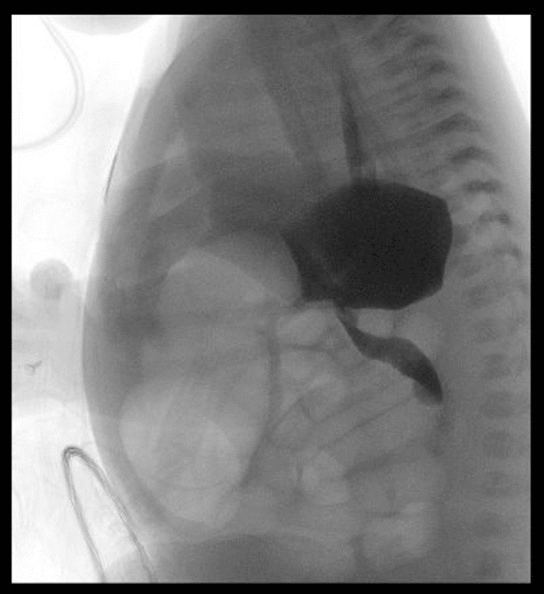
On day of life 1 the baby was tolerating her first breast feeds, however overnight the baby had bilious emesis and had yet to pass meconium. The patient’s abdomen was slightly more distended and tympanic. Plain film showed distended bowel. A replogle tube was placed. Vital signs were within the normal range and labs were unremarkable. Subsequently, an upper GI series was performed and showed a proximal small bowel obstruction.

A contrast enema was done and showed a patent rectum without evidence of a microcolon, Hirchsprung’s disease or malposition. The baby was then taken to the operating room on day of life 2 for an exploratory laparotomy.

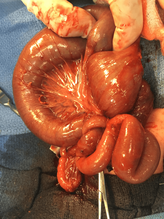
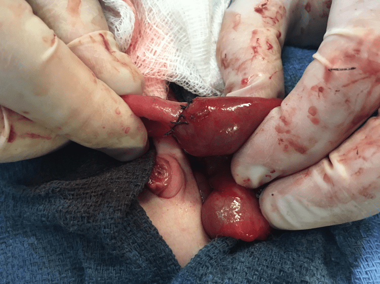
Upon entry into the peritoneal cavity, congenital adhesions were encountered and lysed. A midgut type IIIb intestinal atresia was found with an internal hernia through a mesenteric defect. A 15cm segmental bowel resection was performed with a single layer hand sewn anastomosis was performed. Postoperatively, the baby was extubated and placed on nasal cannula with the replogle in place.

The baby required parental nutrition for weeks after surgery due to inability to consistently tolerate a PO diet despite repeated upper GI films ruling out a mechanical obstruction. Therefore, on post op day 20, the baby started a 5 day course of erythromycin to promote GI motility. After the start of erythromycin, the baby was started on feeds and was gradually advanced with good tolerance. On post op day 37, the baby continue to tolerate a PO diet and was reintroduced to expressed breast milk. The baby was reliably tolerating a home breast feeding schedule by post op day 39. On post op day 41 the baby was discharged home.

Image 1: Antero-Posterior Babygram

Image 2: Left Lateral Decubitus plain film showing air fluid levels and obstruction

Image 3: Upper GI Series demonstrating a proximal intestinal obstruction

Image 4: Contrast Enema demonstrating a patent rectum and colon

Image 5a: Operative photo depicting a Type IIIb intestinal atresia with an internal hernia through a mesenteric defect

Image 5b: Operative photo depicting a Type IIIb intestinal atresia with an internal hernia through a mesenteric defect

Image 6: Operative photo showing a Single Layer hand anastomosis


Presented at the SAGES 2017 Annual Meeting in Houston, TX.

Abstract ID: 94479

Program Number: P644

Presentation Session: Poster Session (Non CME)

Presentation Type: Poster

View this Poster

Related


Hours & Info

15821 Ventura Blvd Ste 400
Encino, CA 91436

1-310-437-0544

[email protected]

Monday – Friday
8am to 5pm Pacific Time

Find Us Around the Web!

  • Bluesky
  • X
  • Instagram
  • Facebook
  • YouTube

Copyright © 2026 · SAGES · All Rights Reserved

Important Links

Healthy Sooner: Patient Information

SAGES Guidelines, Statements, & Standards of Practice

SAGES Manuals

Refine Search