• Skip to main content
  • Skip to header right navigation
  • Skip to site footer

Log in
www.sages.org

SAGES

Reimagining surgical care for a healthier world

  • Home
    • SAGES Home
    • SAGES Foundation Home
  • About
    • Awards
    • Who Is SAGES?
    • Leadership
    • Our Mission
    • Advocacy
    • Committees
      • SAGES Board of Governors
      • Officers and Representatives of the Society
      • Committee Chairs and Co-Chairs
      • Committee Rosters
      • SAGES Past Presidents
    • Why Should You Support SAGES?
    • SAGES Swag
  • Meetings
    • SAGES NBT Innovation Weekend
    • SAGES Annual Meeting
      • 2026 Annual Meeting
      • 2027 Scientific Session Call for Abstracts
      • 2027 Emerging Technology Call for Abstracts
    • CME Claim Form
    • SAGES Past, Present, Future, and Related Meeting Information
    • SAGES Related Meetings & Events Calendar
  • Join SAGES!
    • Membership Application
    • Membership Benefits
    • Membership Types
      • Requirements and Applications for Active Membership in SAGES
      • Requirements and Applications for Affiliate Membership in SAGES
      • Requirements and Applications for Associate Active Membership in SAGES
      • Requirements and Applications for Candidate Membership in SAGES
      • Requirements and Applications for International Membership in SAGES
      • Requirements for Medical Student Membership
    • Member Spotlight
    • Give the Gift of SAGES Membership
  • Patients
    • Join the SAGES Patient Partner Network (PPN)
    • Patient Information Brochures
    • Healthy Sooner – Patient Information for Minimally Invasive Surgery
    • Choosing Wisely – An Initiative of the ABIM Foundation
    • All in the Recovery: Colorectal Cancer Alliance
    • Find A SAGES Surgeon
  • Publications
    • Clinical / Practice / Training Guidelines, Statements, and Standards of Practice
    • Sustainability in Surgical Practice
    • SAGES Stories Podcast
    • SAGES Lead Up Podcast
    • Patient Information Brochures
    • Patient Information From SAGES
    • TAVAC – Technology and Value Assessments
    • Surgical Endoscopy and Other Journal Information
    • Innovative Surgical Trends
    • SAGES Manuals
    • MesSAGES – The SAGES Newsletter
    • COVID-19 Archive
    • Troubleshooting Guides
  • Education
    • Wellness Resources – You Are Not Alone
    • Avoid Opiates After Surgery
    • SAGES Subscription Catalog
    • SAGES TV: Home of SAGES Surgical Videos
    • The SAGES Safe Cholecystectomy Program
    • Masters Program
    • Resident and Fellow Opportunities
      • MIS Fellows Course
      • SAGES Robotics Residents and Fellows Courses
      • SAGES Free Resident Webinar Series
      • Advanced Laparoscopy and Fluorescence-Guided Surgery Course for Fellows
      • Fellows’ Career Development Course
    • SAGES S.M.A.R.T. Enhanced Recovery Program
    • SAGES @ Cine-Med Products
      • SAGES Top 21 Minimally Invasive Procedures Every Practicing Surgeon Should Know
      • SAGES Pearls Step-by-Step
      • SAGES Flexible Endoscopy 101
    • SAGES OR SAFETY Video Activity
    • Foregut Video Atlas
  • Opportunities
    • Join the SAGES Patient Partner Network (PPN)
    • Fellowship Recognition Opportunities
    • SAGES Advanced Flexible Endoscopy Area of Concentrated Training (ACT) SEAL
    • Multi-Society Foregut Fellowship Certification
    • Research Opportunities
    • FLS
    • FES
    • FUSE
    • Jobs Board
    • SAGES Go Global: Global Affairs
  • Learning Hub
You are here: Home / Abstracts / Stereoscopic Augmented Reality Visualization for Laparoscopic Surgery – Initial Clinical Experience

Stereoscopic Augmented Reality Visualization for Laparoscopic Surgery – Initial Clinical Experience

Raj Shekhar, Xinyang Liu, Emmanuel Wilson, Sukryool Kang, Mikael Petrosyan, Timothy D Kane. Children’s National Health System

Objectives: Conventional laparoscopic visualization is often accompanied with a flat representation of the 3D anatomy and limited visualization of structures located beneath visible organ surfaces (e.g., blood vessels, bile ducts, tumors). To address these limitations, we have developed a novel augmented reality (AR) visualization technology that merges real-time laparoscopic ultrasound (LUS) images with live stereoscopic laparoscopic video. This study constitutes initial clinical application of the developed AR system for the purposes of demonstrating technical feasibility and clinical benefits, and obtaining clinically relevant feedback to guide future development.

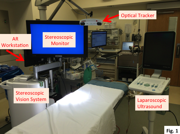
Methods: The AR system includes (1) a stereoscopic vision system for visualizing surgical anatomy with true depth; (2) a laparoscopic ultrasound scanner, which enables visualization of critical internal structures; (3) an optical tracking system, which tracks in real time the two imaging devices by tracking optical markers affixed to custom-designed fixtures; and (4) a computer workstation. With IRB approval, the clinical testing of the AR system in patients undergoing laparoscopic cholecystectomy continues and 7 patients (ages 4-17, 1 male, 6 females) have been recruited to date. Fig. 1 shows the setup of the AR system in the OR. The AR system calibration, performed up to a day in advance, preceded each clinical use. After calibration, the laparoscope, the LUS probe, and the tracking fixtures were sent for sterilization. A specialist assembled the sterilized items in the OR at the beginning of the surgery and attached 4 pre-sterilized optical markers on each of the 2 fixtures. Fig. 2 shows the surgeons, who wear polarized 3D glasses to perceive depth, using the AR system to visualize the hepatobiliary anatomy. The AR visualization was performed for up to 5 min prior to starting the actual surgery. When instructed, the system records live LUS images, stereoscopic laparoscopic images, and stereoscopic AR images as digital videos.

imageimage

Results: The ongoing clinical testing has been a learning process for the project team to successfully use the AR system in the OR. During the first 3 cases, several technical and practical issues prevented obtaining well-registered AR images. With these issues resolved, the AR system has worked successfully in the following 4 cases. On average, the system took 15 min to set up in the OR, and this setup took place in parallel with patient setup in conventional laparoscopic surgery. The use of the AR system added approximately 5 min to the clinical procedure. The AR system was considered easy to use and helpful in the visualization of normally unseen structures. Fig. 3 shows the system’s ability to visualize the hepatic artery and the cystic duct in the conduct of laparoscopic cholecystectomy. The system was also used to visualize the hepatic vasculature and the pancreatic duct.

image

Conclusions: We have successfully translated the AR system from the laboratory to the operating room. The transformative clinical application envisioned for the use of this technology will be in aiding visualization and accurate resection of subparenchymal lesions (liver, kidney, lung) by minimally invasive approaches.

Related


Hours & Info

15821 Ventura Blvd Ste 400
Encino, CA 91436

1-310-437-0544

[email protected]

Monday – Friday
8am to 5pm Pacific Time

Find Us Around the Web!

  • Bluesky
  • X
  • Instagram
  • Facebook
  • YouTube

Copyright © 2026 · SAGES · All Rights Reserved

Important Links

Healthy Sooner: Patient Information

SAGES Guidelines, Statements, & Standards of Practice

SAGES Manuals

Refine Search