Venkata S Kanthimathinathan, MD, Camila Manrique Acevedo, MD, Arthur Rawlings, MD. University of Missouri Health Care
Introduction:
Diagnosis of bilateral adrenal mass in any patient presents a management dilemma. We report an unusual case of a 51 year old female with 4.8 cm right adrenal pheochromocytoma and a 3 mm left adrenal lesion. Despite extensive work-up including blood, urine, CT scan, MRI, MIBG scan, we were unable to reach a definitive diagnosis on the left adrenal lesion. We performed laparoscopic right adrenalectomy for the right adrenal tumor. One month postoperative blood and urine work up revealed normal metanephrines. As there is no evidence of hormonally active tumor, we are observing the left adrenal lesion with biochemical and radiological investigations.
Case report:
51 year old Caucasian female with history of ulcerative colitis, referred to General surgery for evaluation of enlarging right adrenal mass. Mass was detected 6 years ago through CT scan done for GI symptoms. No further follow-up was done until this time. Patient reports intermittent bilateral flank pain, night sweats, and palpitations. Physical examination was unremarkable.
Pertinent laboratory values include elevated urine metanephrine (1555), urine normetanephrine (262), serum normetanephrine (1.22) and serum metanephrine (5.82). She has normal aldosterone, renin, ACTH, DHEA and 24 hour urine cortisol levels.
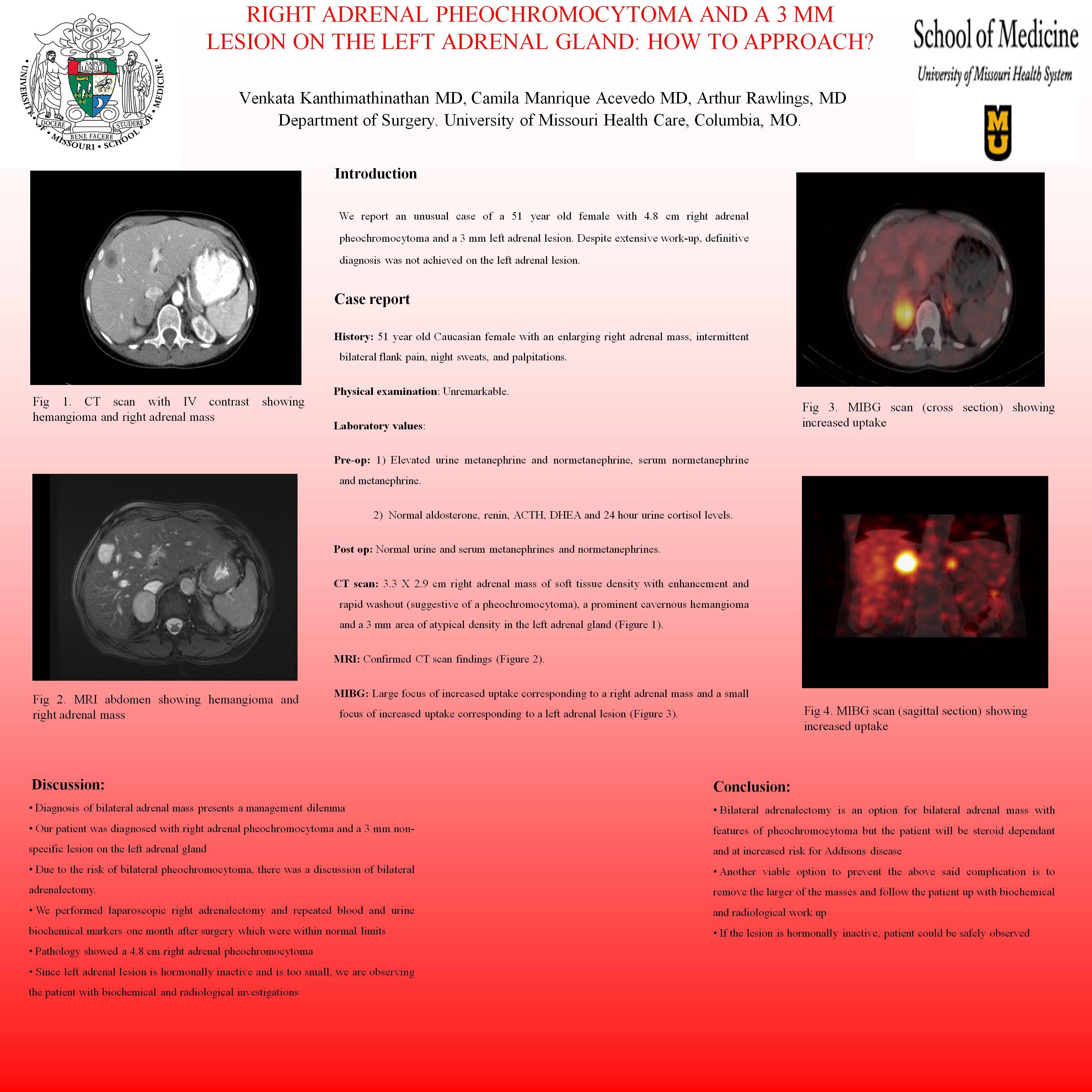
Radiological investigations include CT scan of the abdomen and pelvis with IV contrast, MRI abdomen and MIBG scintigraphy with SPECT/CT scan. CT scan showed a 3.3 X 2.9 cm right adrenal mass that appears to be of soft tissue density with enhancement and rapid washout, suggestive of a pheochromocytoma. CT also showed a prominent cavernous hemangioma and a very subtle area of atypical density in the left adrenal gland that measures approximately 3 mm in size, relatively nonspecific in appearance. MRI confirmed CT scan findings. MIBG scan showed a large focus of increased uptake corresponding to a right adrenal mass on SPECT CT images and a small focus of increased uptake corresponding to the normal sized left adrenal gland.
Patient was diagnosed with right adrenal pheochromocytoma and a non specific 3 mm lesion in the left adrenal gland. As the lesion on the left adrenal gland was too small to characterize, we performed a laparoscopic right adrenalectomy. Her surgery and postoperative course were uneventful and she was discharged on postoperative day 2. Pathology showed a 4.8 cm right adrenal pheochromocytoma with margins free of tumor. Repeat blood and urine work up one month after surgery revealed normal urine metanephrine, normetanephrine, serum metanephrine and normetanephrine. However, genetic testing returned SDHB mutation positive putting the patient at high risk for malignant disease and extra-adrenal tumors. As there is no evidence of hormonally active tumor, we are currently observing the left adrenal lesion with biochemical and radiological investigations.
Session Number: Poster – Poster Presentations
Program Number: P603
View Poster