Nobumi Tagaya, PhD, Yawara Kubota, MD, Asami Suzuki, MD, Nana Makino, MD, Kosuke Hirano, MD, Shinichiro Kouketsu, PhD, Emiko Takeshita, PhD, Yoshitake Sugamata, PhD, Hidemaro Yoshiba, PhD, Shinichi Sameshima, PhD, Masatoshi Oya, PhD. First Department of Surgery, Dokkyo Medical University Koshigaya Hospital
Background: We evaluate the real-time fluorescence imaging of biliary anatomy using indocyanine green (ICG) during single-incision laparoscopic cholecystectomy.
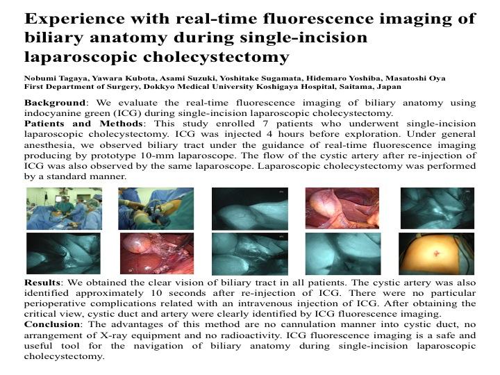
Patients and Methods: This study enrolled 7 patients who underwent single-incision laparoscopic cholecystectomy. ICG was injected 4 hours before exploration. Under general anesthesia, we observed biliary tract under the guidance of real-time fluorescence imaging producing by prototype 10-mm laparoscope. The flow of the cystic artery after re-injection of ICG was also observed by the same laparoscope. Laparoscopic cholecystectomy was performed by a standard manner
Results: We obtained the clear vision of biliary tract in all patients. The cystic artery was also identified approximately 10 seconds after re-injection of ICG. There were no particular perioperative complications related with an intravenous injection of ICG. After obtaining the critical view, cystic duct and artery were clearly identified by ICG fluorescence imaging.
Conclusion: The advantages of this method are no cannulation manner into cystic duct, no arrangement of X-ray equipment and no radioactivity. ICG fluorescence imaging is a safe and useful tool for the navigation of biliary anatomy during single-incision laparoscopic cholecystectomy.
Session Number: Poster – Poster Presentations
Program Number: P327
View Poster