Objectives: Natural Orifice Translumenal Endoscopic Surgery (NOTES) utilizes resectional or anastomotic techniques. Anastomotic work to date is limited to suturing and stapling devices. Endoscopic gastrojejunostomy has been reported in two animal studies. The aim of our study is to assess feasibility of gastrojejunostomy with full thickness plications.
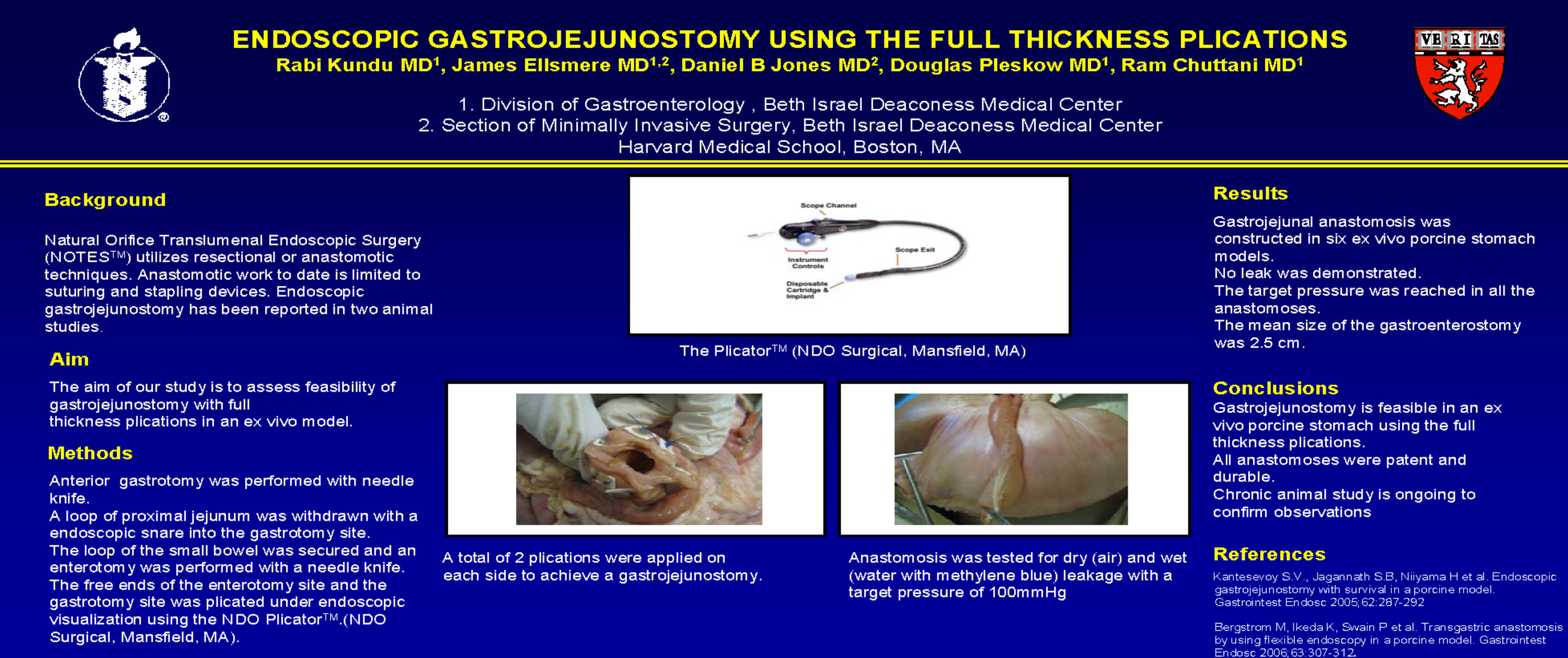
Methods: Six ex vivo porcine stomachs were prepared with small bowel in continuity. Using an end viewing endoscope, transillumination and palpation were used to select the site of the anterior gastrotomy. The gastrotomy was performed with needle knife and was extended with a traction sphincterotome. A loop of proximal jejunum was withdrawn with a endoscopic snare into the gastrotomy site. Once the loop of the small bowel was secured an enterotomy was performed with a needle knife and sphincterotome using cautery or a new technique with a snare. The free ends of the enterotomy site and the gastrotomy site was plicated under endoscopic visualization using the NDO PlicatorTM.(NDO Surgical, Mansfield, MA). A total of 2 plications were applied on each side to achieve a gastrojejunostomy. Anastomosis was tested for dry (air) and wet (water with methylene blue) leakage with a target pressure of 100mmHg.
Results: Gastrojejunal anastomosis was constructed in six ex vivo porcine stomach models. No leak was demonstrated. The target pressure was reached in all the anastomoses. The mean size of the gastroenterostomy was 2.5 cm.
Conclusions: Gastroenterostomy is feasible in an ex vivo porcine stomach using the full thickness plications. All anastomoses were patent and durable. Chronic animal study is ongoing to confirm observations.
Session: Poster
Program Number: P223
