Katsunori Nishikawa, MD, Noburo Omura, MD, Masami Yuda, MD, Yujiro Tanaka, MD, Akira Matsumoto, MD, Yuichiro Tanishima, MD, Toshiyuki Sasaki, MD, Yoshiro Ishibashi, MD, Kouji Nakada, MD, Norio Mitsumori, MD, Hideyuki Kashiwagi, MD, Katsuhiko Yanaga, MD. The Jikei University School of Medicine Department of Surgery
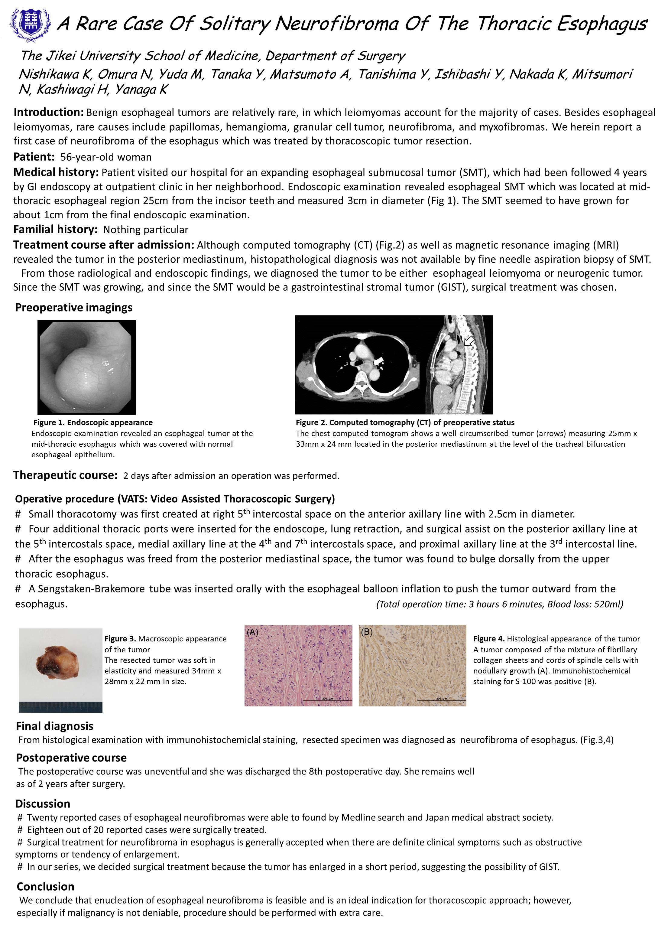
Benign esophageal tumors are rare disease, of which leiomyomas account for the majority of disease, while uncommon tumors include papilloma(s), hemangioma, granular cell tumor, neurofibroma, and myxofibroma(s). We present a case of a 56-year-old woman with the diagnosis of esophageal submucosal tumor (SMT) in mid-portion of the thoracic esophagus. The SMT was 3 cm in diameter, and MRI suggested the diagnosis of esophageal leiomyoma or neurofibroma. The operation was performed by video-assisted thoracoscopic surgery (VATS). In order to protrude SMT outward from the esophagus for easy access, a Sengstaken-Brakemore tube was inflated in the esophageal lumen at the level of the tumor. The tumor was gently enucleated from the esophageal wall by splitting esophageal muscle layers without mucosal injury. Her postoperative course was uneventful and the patient was discharged on postoperative day 8, and she remains asymptomatic to date. Histrogical diagnosis of the tumor was esophageal neurofibroma. To our knowledge, this is the first report of such a tumor in the esophagus treated by VATS.
Session Number: Poster – Poster Presentations
Program Number: P531
View Poster