Vadim P Koshenkov, MD, Zoltan H Nemeth, MD PhD, Jain Joseph, MD, Mitchel S Carter, MD, Alexander Abkin, MD. Morristown Memorial Hospital, Atlantic Health
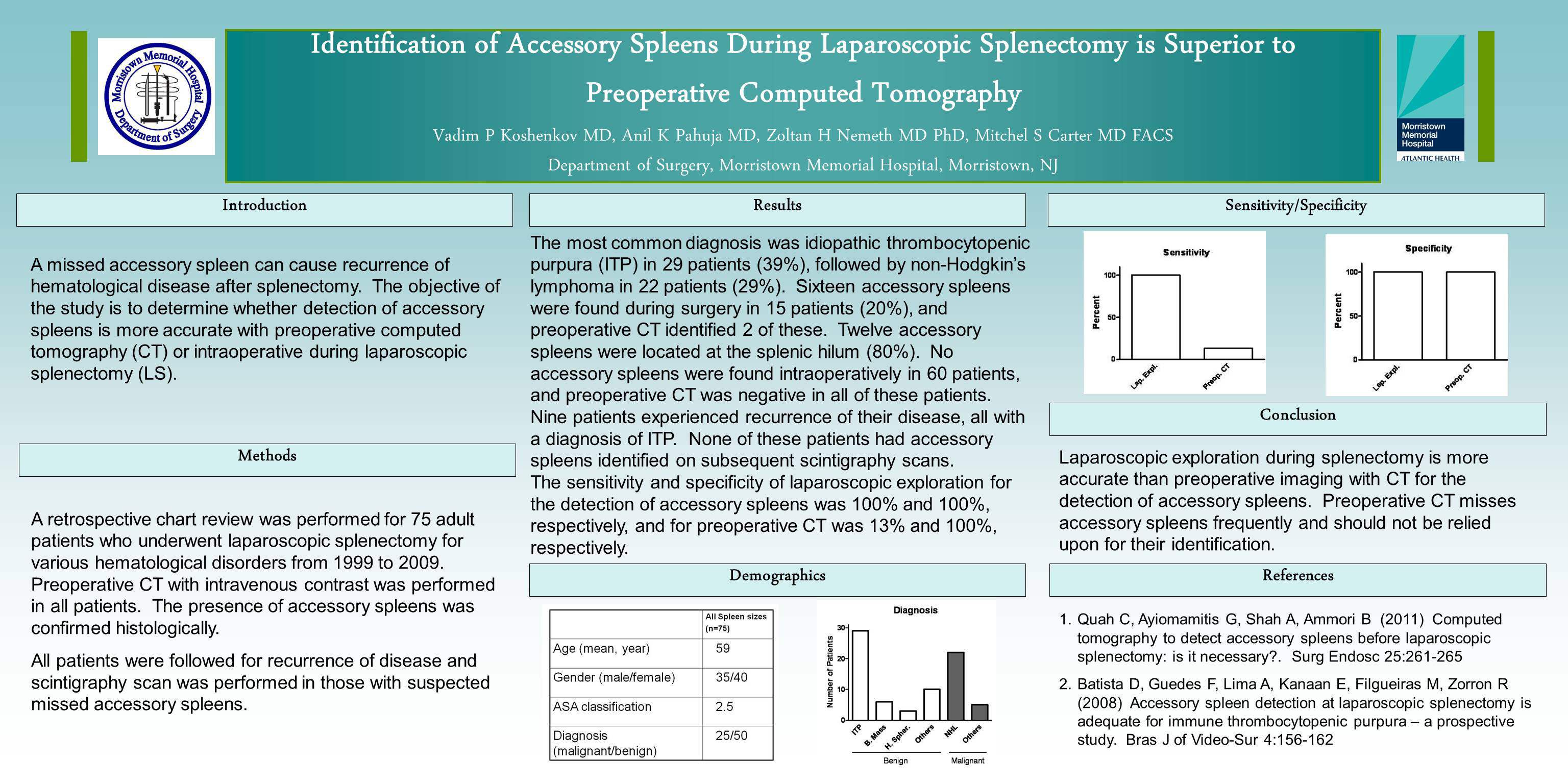
INTRODUCTION: A missed accessory spleen can cause recurrence of hematological disease after splenectomy. The objective of the study is to determine whether the detection of accessory spleens is more accurate with preoperative computed tomography (CT) or intraoperative exploration during laparoscopic splenectomy (LS).
METHODS: A retrospective chart review was performed for 75 adult patients who underwent laparoscopic splenectomy for various hematological disorders from 1999 to 2009. Preoperative CT with intravenous contrast was done in all patients. Presence of accessory spleens was confirmed histologically. All patients were followed for recurrence of disease and scintigraphy scan was performed in those with suspected missed accessory spleens.
RESULTS: The most common diagnosis was idiopathic thrombocytopenic purpura (ITP) in 29 patients (39%), followed by non-Hodgkin’s lymphoma in 22 patients (29%). Sixteen accessory spleens were found during surgery in 15 patients (20%), and preoperative CT identified 2 of these. Twelve accessory spleens were located at the splenic hilum (80%). No accessory spleens were found intraoperatively in 60 patients, and preoperative CT was negative in all of them. Nine patients experienced recurrence of their disease, all with diagnosis of ITP. None of these patients had accessory spleens identified on subsequent scintigraphy scans. The sensitivity and specificity of laparoscopic exploration for the detection of accessory spleens was 100% and 100%, respectively, and of the preoperative CT was 13% and 100%, respectively.
CONCLUSIONS: Laparoscopic exploration during splenectomy is more accurate than preoperative imaging with CT for the detection of accessory spleens. Preoperative CT misses accessory spleens frequently and should not be relied upon for their identification.
Session: Poster
Program Number: P508
View Poster