Introduction: Appendectomy and small bowel enterolysis for adhesions are commonly performed general surgical procedures. Our lab aimed to assess the feasibility of natural orifice translumenal endoscopic surgery (NOTES) for appendectomy and lysis of adhesions (LOA) using a live porcine model.
Methods: The bicornuate uterus of the female swine has been used previously as a model for appendectomy in laparoscopic procedures. In this non-survival study, 15 uterine horn resections were attempted in 11 swine. In five of these animals, 3-4 simulated small bowel adhesions (15 total) were created by laparoscopically suturing the bowel to the abdominal wall. In all cases, a dual channel endoscope was used to create an endoscopic gastrotomy to gain access to the abdominal cavity. A ShapeLock endoscopic guide was also used to assist with navigation of the endoscope within the abdomen. In the swine with simulated adhesions, LOA was performed first using endoscopic shears by an operator blinded to the locations of the adhesions. Uterine horn dissection was accomplished using standard endoscopic needle knife cautery. A Carter-Thomason 2-mm needle point suture passer was percutaneously introduced to assist with uterine horn retraction. This was the only laparoscopic instrument used during the procedures. After mobilization of the uterine horn, the stump was ligated with either clips or a detatchable loop ligating device. The uterine horn was then divided and withdrawn through the stomach.
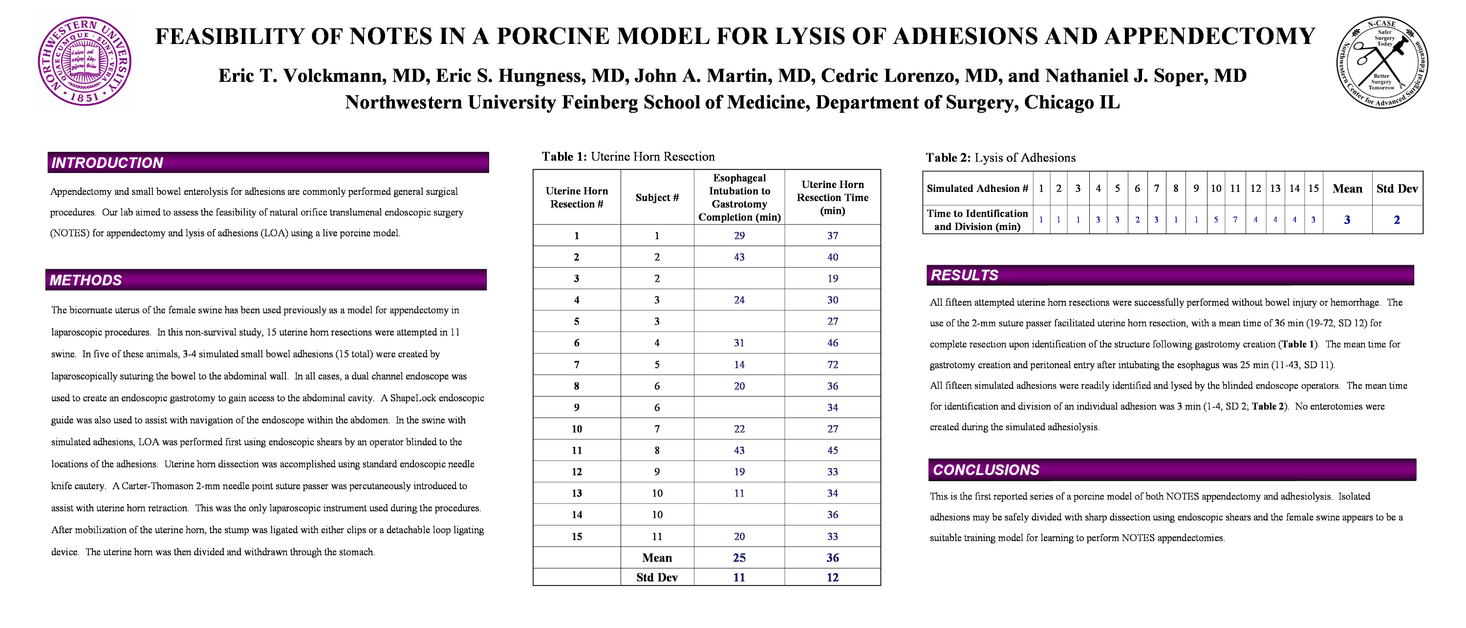
Results: All fifteen simulated adhesions were readily identified and lysed by the blinded endoscope operators. The mean time for identification and division of an individual adhesion was 2 min (1-4, SD 2). No enterotomies were created during the simulated adhesiolysis. All fifteen attempted uterine horn resections were successfully performed without bowel injury or hemorrhage. The use of the 2-mm suture passer facilitated uterine horn resection, with a mean time of 36 min (19-72, SD 12) for complete resection upon identification of the structure following gastrotomy creation. The mean time for gastrotomy creation and peritoneal entry after intubating the esophagus was 25 min (11-43, SD 11)
Conclusion: This is the first reported series of a porcine model of both NOTES appendectomy and adhesiolysis. Isolated adhesions may be safely divided with sharp dissection using endoscopic shears and the female swine appears to be a suitable training model for learning to perform NOTES appendectomies.
Session: Poster
Program Number: P210
